. 2012 Jan 3:2:102.
doi: 10.3389/fendo.2011.00102. eCollection 2011.

β-Adrenoceptor Signaling Networks in Adipocytes for Recruiting Stored Fat and Energy Expenditure

Affiliations

β-Adrenoceptor Signaling Networks in Adipocytes for Recruiting Stored Fat and Energy Expenditure

Sheila Collins. Front Endocrinol (Lausanne). .

Abstract

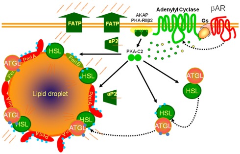
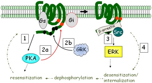
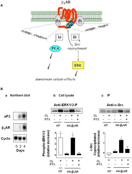
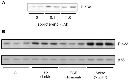
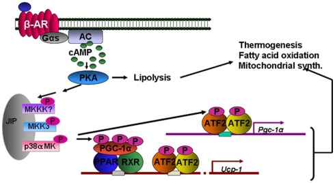
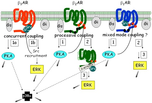
THE ADIPOCYTE IS LIKE A BANK: a place to store excess (caloric) cash in times of plenty, and from which one can withdraw savings during "lean times." The β-adrenoceptors (βAR) are the gateways to this mobilization of fat to be consumed in other tissues. This review discusses the βAR signaling pathway(s) in white and brown adipocytes. Studies in rodent models show that brown adipocytes nestled with white fat depots correlate with and are considered a key enabling factor in resistance to diet-induced obesity. Since it is now recognized that adult humans have brown adipocytes, knowing the steps in these signaling pathways may provide the opportunity to manipulate adipocytes to be net consumers of energy.

Keywords: adipocyte; adrenergic; brown; kinase; signaling; white.


Figures

Figure 1
Figure 1
Scheme for βAR signaling cascades controlling lipolysis in adipocytes. βARs activate adenylyl cyclase through their coupling to the heterotrimeric Gs, producing cAMP (green dots) from ATP (yellow dots) to activate the cAMP-dependent protein kinase (PKA), allowing the catalytic subunits (PKA-C2) to be released from the holoenzyme that is anchored to the plasma membrane (AKAP). PKA phosphorylates (blue dots) lipases (HSL: hormone sensitive lipase) and lipid droplet binding proteins such as perilipins (Peri A; Peri B). Adipose triglyceride lipase (ATGL) is phosphorylated but not by PKA. The fatty acids released from triglyceride are chaperoned out of the cell by lipid binding proteins (aP2) and exported through fatty acid transport proteins (FATP).
Figure 2
Figure 2
βARs activate ERK MAPK in addition to PKA: the β2AR mechanism as an example. Upon catecholamine activation, PKA is activated (step 1), and phosphorylates the receptor at intracellular sites (red circles; step 2a). The receptor is also phosphorylated by G protein-coupled receptor kinase (GRK) at multiple sites in the C-terminus (step 2b). PKA phosphorylation interdicts interaction with Gs to favor Gi, while GRK recruits β-arrestin, promoting ERK activation (step 3). β2AR is subject to rapid desensitization and internalization (step 4), whereupon phosphatases remove the phosphates and the receptor is recycled back to the plasma membrane.
Figure 3
Figure 3
A unique dual signaling mechanism by β3AR. (A) The receptor can couple to both Gs and Gi to activate PKA (step 1a) and ERK (step 1b). The shaded gray arrows point to subsequent downstream cellular effects. (B) Preadipocytes were transfected or not (NT) with hemagglutinin (HA)-tagged β3AR and differentiated. (a) Expression of aP2 and β3AR mRNA as a function of differentiation on the indicated days. Levels of aP2 are maximal by day 4. Cyclophilin RNA (Cyclo) is the internal control. (b) Phospho-ERK1/2 in cell lysates in response to the β3AR agonist CL316243 (CL) in the absence or presence of pertussis toxin (PTX; n = 3; mean ± SD). (c) Level of c-Src co-precipitated with β3AR (n = 3; mean ± SD). (Adapted from Cao et al., 2000).
Figure 4
Figure 4
β-agonists increase activation of p38 MAPK in cultured human subcutaneous adipocytes. (A) Western blot measuring isoproterenol (Iso) concentration-response for phospho-p38 MAPK (P-p38). (B) Western blot comparing P-p38 response to 15 min treatment with Iso, EGF and the positive control cell stressor Anisomycin. All samples shown are independent biological replicates.
Figure 5
Figure 5
βAR activation of thermogenesis through p38 MAPK. Direct targets of p38 MAPK include peroxisome proliferator-activated receptor-gamma coactivator-1α (PGC-1α) and ATF-2. These target the genes for Pgc-1α and Ucp1. The tandem arrows between PKA and the MAPK module are not yet biochemically defined.
Figure 6
Figure 6
Signaling networks of the βARs in adipocytes. Each of the three βARs is able to activate similar pathways: PKA, ERK, and p38 MAPK. However the order and number of steps involved varies, such that β3AR represents what we term “concurrent” coupling to Gs vs. Gi, vs. the β2AR type of “processive” coupling. Less is known about the details of β1AR. Because β1AR also contains one of the PXXP motifs as in β3AR, but it is also phosphorylated, we have provisionally termed in “mixed-mode” coupling. The shaded gray arrows point to subsequent downstream cellular effects.

References

    1. Arch J. R. (2002). beta(3)-Adrenoceptor agonists: potential, pitfalls and progress. Eur. J. Pharmacol. 440, 99–10710.1016/S0014-2999(02)01421-8 - DOI - PubMed
    1. Arch J. R. S., Ainsworth A. T., Ellis R. D., Piercy V., Thody V. E., Thurlby P. L., Wilson C., Wilson S., Young P. (1984). Treatment of obesity with thermogenic β-adrenoceptor agonists: studies on BRL 28630A in rodents. Int. J. Obes. 8, 1–11 - PubMed
    1. Bachman E. S., Dhillon H., Zhang C. Y., Cinti S., Bianco A. C., Kobilka B. K., Lowell B. B. (2002). betaAR signaling required for diet-induced thermogenesis and obesity resistance. Science 297, 843–84510.1126/science.1073160 - DOI - PubMed
    1. Bickel P. E., Tansey J. T., Welte M. A. (2009). PAT proteins, an ancient family of lipid droplet proteins that regulate cellular lipid stores. Biochim. Biophys. Acta 1791, 419–440 - PMC - PubMed
    1. Brasaemle D. L. (2007). Thematic review series: adipocyte biology. The perilipin family of structural lipid droplet proteins: stabilization of lipid droplets and control of lipolysis. J. Lipid Res. 48, 2547–255910.1194/jlr.R700014-JLR200 - DOI - PubMed

LinkOut - more resources