. 2012 Mar 2:3:38.
doi: 10.3389/fendo.2012.00038. eCollection 2012.

Insulin-like growth factor binding proteins: a structural perspective

Affiliations

Insulin-like growth factor binding proteins: a structural perspective

Briony E Forbes et al. Front Endocrinol (Lausanne). .

Abstract

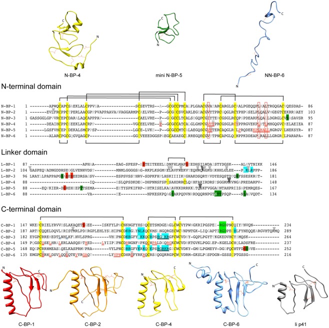
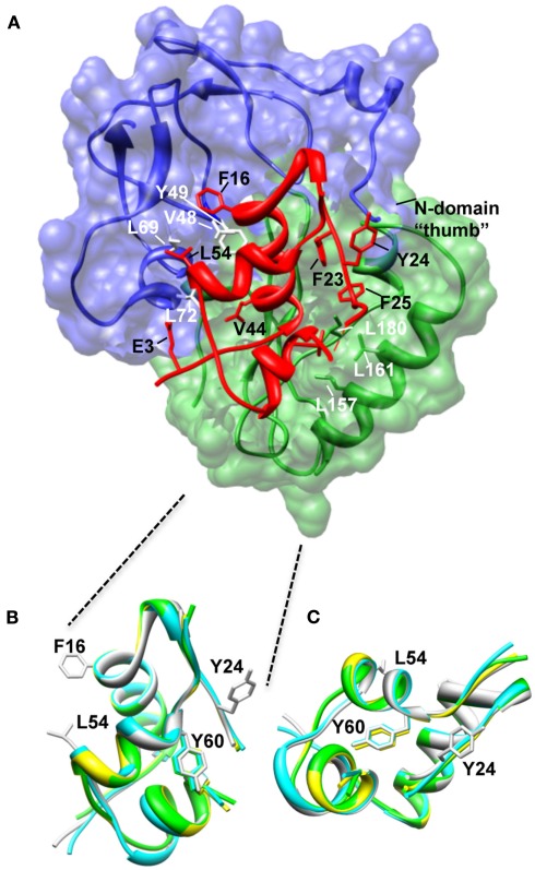
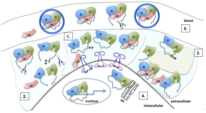
Insulin-like growth factor binding proteins (IGFBP-1 to -6) bind insulin-like growth factors-I and -II (IGF-I and IGF-II) with high affinity. These binding proteins maintain IGFs in the circulation and direct them to target tissues, where they promote cell growth, proliferation, differentiation, and survival via the type 1 IGF receptor. IGFBPs also interact with many other molecules, which not only influence their modulation of IGF action but also mediate IGF-independent activities that regulate processes such as cell migration and apoptosis by modulating gene transcription. IGFBPs-1 to -6 are structurally similar proteins consisting of three distinct domains, N-terminal, linker, and C-terminal. There have been major advances in our understanding of IGFBP structure in the last decade and a half. While there is still no structure of an intact IGFBP, several structures of individual N- and C-domains have been solved. The structure of a complex of N-BP-4:IGF-I:C-BP-4 has also been solved, providing a detailed picture of the structural features of the IGF binding site and the mechanism of binding. Structural studies have also identified features important for interaction with extracellular matrix components and integrins. This review summarizes structural studies reported so far and highlights features important for binding not only IGF but also other partners. We also highlight future directions in which structural studies will add to our knowledge of the role played by the IGFBP family in normal growth and development, as well as in disease.

Keywords: IGF binding protein; insulin-like growth factor; protein structure.


Figures

Figure 1
Figure 1
Sequences and structures of IGFBP domains. Sequence alignments of IGFBP-1 to-6 N-terminal (N-), linker (L-), and C-terminal (C-) domains, highlighting cysteine residues (yellow box) and the disulfide bonding connectivity (black lines above and below sequences). Residue numbers are shown next to the sequences and every 10th residues is gray or white. The following distinctive features are highlighted: Red underlined text highlights IGF binding residues defined in structural studies of the N-domain [IGFBP-4 (Sitar et al., 2006) and mini IGFBP-5 (Kalus et al., 1998)] and C-domain [IGFBP-4 (Sitar et al., 2006) and IGFBP-6 (Headey et al., 2004a)]. Open red boxes, key residues involved in binding conserved across all IGFBPs; Light gray arrowheads, IGFBP-2 proteolysis sites (Ho and Baxter, ; Rehault et al., ; Monget et al., ; Standker et al., ; Mark et al., ; Berg et al., ; Miyamoto et al., 2007); Dark gray arrowhead, PAPP-A cleavage sites on IGFBP-2, -4, and -5 (Conover et al., ; Laursen et al., ; Monget et al., 2003); Blue boxes, IGFBP heparin-binding domains confirmed by site-directed mutagenesis or NMR-IGFBP-2 (Russo et al., ; Kuang et al., 2006), IGFBP-3 (Firth et al., 1998), and IGFBP-5 (Arai et al., 1996b); Filled red boxes, phosphorylation sites (Jones et al., ; Coverley et al., ; Gibson et al., ; Graham et al., ; Dolcini et al., 2009); Light green boxes, integrin binding sites (Jones et al., ; Kuang et al., 2006), Dark green boxes; glycosylation sites (Neumann et al., ; Firth and Baxter, ; Graham et al., 2007); Black underlined text, nuclear localization sequences (Schedlich et al., ; Iosef et al., 2008); Blue underlined text, Leu194 and Leu197 of IGFBP-3 involved in nuclear export (Paharkova-Vatchkova and Lee, 2010). Protein sequence SwissProt accession numbers are as follows: IGFBP-1 (P08833), IGFBP-2 (P18065), IGFBP-3 (P17936), IGFBP-4 (P22692), IGFBP-5 (P24593), IGFBP-6 (P24592). N-domain and C-domain ribbon structures are from the following database files: N-BP-4 (PDB 2DSR), mini N-BP-5 (PDB 1BOE, conformer 1), NN-BP-6 (PDB 2JM2), C-BP-1 (PDB 1ZT3), C-BP-2 (PDB 2H7T), C-BP-4 (PDB 2DSR), C-BP-6 (PDB 1RMJ), and the Ii p41 thryglobulin type 1 domain (PDB 1ICF). Some of these structures are derived from IGF:IGFBP complexes (see Table 1). For those NMR PDB files containing more than one conformer the first entry was used in this figure. Molecular graphic images were produced using UCSF Chimera program from Resource for Biocomputing, Visualization, and Informatics at the University of California at San Francisco (http://www.cgl.ucsf.edu/chimera; Pettersen et al., 2004).
Figure 2
Figure 2
Structure of the N-BP-4:IGF-I:C-BP-4 complex (PDB 2DSR; Sitar et al., 2006). (A) The N-domain of BP-4 (blue, residues 3–82) contacts the C-domain of BP-4 (green, residues 151–232) to form a high-affinity complex with IGF-I (red). IGF-I residues Phe16 and Leu54 insert into a cleft in the N-BP-4 encompassing residues Val48, Tyr49, Leu69, and Leu72 (Kalus et al., 1998). The “thumb” region of the N-domain and residues Leu157, Leu161, and Ile180 of the IGFBP-4 C-domain are in contact with IGF-I residues Phe23, Tyr24, Phe25, and Val44, which are involved in IGF-1R binding, resulting in steric hindrance of IGF-I binding to the receptor (PDB 2DSR, Sitar et al., 2006). (B,C) Superposition of IGF-I crystal structures highlighting that the Tyr60 side chain remains in the same position whether IGF-I is bound to IGFBPs or free. Ribbon views of IGF-I in the free form (bound to detergent molecules) PDB 1GZR (green; Brzozowski et al., 2002) and PDB 1IMX (yellow; Vajdos et al., 2001), the IGF-I‚ mini-N-BP-5 binary complex (PDB 1H59, blue; Zeslawski et al., 2001), and the IGF-I‚ N-BP-4‚ C-BP-4 ternary complex (PDB 2DSR, light gray; Sitar et al., 2006) are superimposed over backbone heavy atoms of the three helices (Ala8–Cys18, Gly42–Cys48, Leu54–Cys61). The side chains of Phe16, Phe49, and Leu54 are shown. The structures are shown in two different orientations (B,C). The structures in (B) are in an orientation equivalent to that of the IGF-I structure in (A). Parts of the flexible regions (residues 1–2, 26–42, and 63–70) are excluded for clarity. Molecular graphic images were produced with the UCSF Chimera program from Resource for Biocomputing, Visualization, and Informatics at the University of California at San Francisco.
Figure 3
Figure 3
Model of the molecular mechanisms of IGFBP action. 1. The N- (blue) and C- (green) domains are connected by a flexible linker (dark blue). Inter-domain interaction between the N- and C-domains is detected in the absence of ligand, but this is of low affinity (Kuang et al., 2007). The interface between the N- and C-domains lies immediately adjacent to their IGF binding surfaces and both domains act cooperatively to bind IGF. Upon IGF-I or IGF-II (pink) binding, the C-domain undergoes a structural change. Residues in the N- and C-domains form the IGF binding site, with the linker playing an as yet unknown role. 2. Proteolysis of IGFBPs occurs primarily in the linker domain (indicated by scissors) resulting in an increased concentration of free IGFs. Individual N- and C-domains have low affinity for IGFs, whereas in vitro, reconstituted binary complexes of the N- and C-domains generally bind IGFs with an affinity only 10-fold lower than intact IGFBPs. It is not clear whether in vivo cleavage results in the dissociation of IGFBP into separate domains or whether cleaved IGFBP domains remain associated via the inter-domain interface. Cleavage products are susceptible to further breakdown, which possibly leads to the equilibrium being pushed toward free IGF and individual IGFBP domains. 3. Intact and cleaved IGFBPs bind extracellular matrix components (ECM, glycosaminoglycans, and proteins) via the C-domain (and the linker domain in IGFBP-2). Binding to ECM (hatched green shape) lowers the affinity for IGFs. 4. IGFBP-2, -3, -5, and -6 are found within the nucleus and can affect gene expression (arrow). IGFBP-3 can be associated with the endoplasmic reticulum (ER). 5. Binary IGFBP:IGF complexes, intact IGFBPs, and cleavage products can be found in the blood. IGFBP-3 and -5 form a trimeric 150 kDa complex with IGF and the acid labile subunit (ALS, large blue circle). Even with limited proteolysis in the L-domain, IGFBP fragments can also form a trimeric complex. Only the binary complexes are small enough to cross the vascular epithelium into the extracellular space to promote IGF-dependent biological activities.

References

    1. Arai T., Busby W., Jr., Clemmons D. R. (1996a). Binding of insulin-like growth factor (IGF) I or II to IGF-binding protein-2 enables it to bind to heparin and extracellular matrix. Endocrinology 137, 4571–457510.1210/en.137.11.4571 - DOI - PubMed
    1. Arai T., Clarke J., Parker A., Busby W., Jr., Nam T., Clemmons D. R. (1996b). Substitution of specific amino acids in insulin-like growth factor (IGF) binding protein 5 alters heparin binding and its change in affinity for IGF-I response to heparin. J. Biol. Chem. 271, 6099–610610.1074/jbc.271.11.6099 - DOI - PubMed
    1. Arai T., Parker A., Busby W., Jr., Clemmons D. R. (1994). Heparin, heparan sulfate, and dermatan sulfate regulate formation of the insulin-like growth factor-I and insulin-like growth factor-binding protein complexes. J. Biol. Chem. 269, 20388–20393 - PubMed
    1. Azar W. J., Azar S. H., Higgins S., Hu J. F., Hoffman A. R., Newgreen D. F., Werther G. A., Russo V. C. (2011). IGFBP-2 enhances VEGF gene promoter activity and consequent promotion of angiogenesis by neuroblastoma cells. Endocrinology 152, 3332–334210.1210/en.2011-1121 - DOI - PubMed
    1. Bach L. A., Headey S. J., Norton R. S. (2005). IGF-binding proteins – the pieces are falling into place. Trends Endocrinol. Metab. 16, 228–23410.1016/j.tem.2005.05.005 - DOI - PubMed

LinkOut - more resources