. 2012 May 25:3:119.
doi: 10.3389/fimmu.2012.00119. eCollection 2012.

Mast cell chemotaxis - chemoattractants and signaling pathways

Affiliations

Mast cell chemotaxis - chemoattractants and signaling pathways

Ivana Halova et al. Front Immunol. .

Abstract

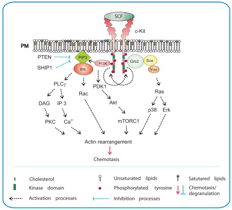
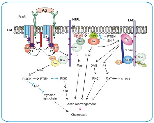
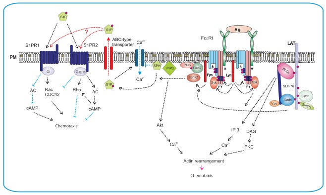
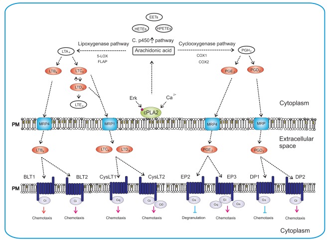
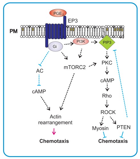
Migration of mast cells is essential for their recruitment within target tissues where they play an important role in innate and adaptive immune responses. These processes rely on the ability of mast cells to recognize appropriate chemotactic stimuli and react to them by a chemotactic response. Another level of intercellular communication is attained by production of chemoattractants by activated mast cells, which results in accumulation of mast cells and other hematopoietic cells at the sites of inflammation. Mast cells express numerous surface receptors for various ligands with properties of potent chemoattractants. They include the stem cell factor (SCF) recognized by c-Kit, antigen, which binds to immunoglobulin E (IgE) anchored to the high affinity IgE receptor (FcεRI), highly cytokinergic (HC) IgE recognized by FcεRI, lipid mediator sphingosine-1-phosphate (S1P), which binds to G protein-coupled receptors (GPCRs). Other large groups of chemoattractants are eicosanoids [prostaglandin E(2) and D(2), leukotriene (LT) B(4), LTD(4), and LTC(4), and others] and chemokines (CC, CXC, C, and CX3C), which also bind to various GPCRs. Further noteworthy chemoattractants are isoforms of transforming growth factor (TGF) β1-3, which are sensitively recognized by TGF-β serine/threonine type I and II β receptors, adenosine, C1q, C3a, and C5a components of the complement, 5-hydroxytryptamine, neuroendocrine peptide catestatin, tumor necrosis factor-α, and others. Here we discuss the major types of chemoattractants recognized by mast cells, their target receptors, as well as signaling pathways they utilize. We also briefly deal with methods used for studies of mast cell chemotaxis and with ways of how these studies profited from the results obtained in other cellular systems.

Keywords: IgE receptor; cell migration; chemoattractant; chemotaxis; mast cell; plasma membrane; signal transduction.


Figures

Figure 1
Figure 1
SCF-mediated chemotaxis. Crosslinking of the plasma membrane (PM) anchored c-Kit by SCF results in dimerization of the receptor and its auto-transphosphorylation. This is followed by recruitment of SH2-domain containing proteins such as PI3K and Grb2 to c-Kit. Activated PI3K generates PIP3, a binding site for PH-domain containing protein Btk, and facilitates further propagation of the signal through activation of PLCγ. An increased activity of PLCγ leads to production of DAG and IP3 and release of Ca2+. This is followed by actin rearrangements and chemotactic response. Production of PIP3 also leads to recruitment of phosphoinositide-dependent kinase (PDK)-1 and Akt to the plasma membrane and subsequent phosphorylation of Akt by PDK1. Akt directly phosphorylates the negative regulator of mTORC1, inactivating thereby its inhibitory action. PIP3 levels are negatively regulated by phosphatases PTEN and SHIP1. Grb2 orchestrates activation of Ras by recruiting Ras and Rho family GEFs, Sos, and Vav. Through Ras, both p38 and Erk are activated and chemotaxis is also promoted in this way. Signaling molecules in bodies are those located at their sites of action and/or bound to the indicated target molecules; other signaling molecules are presented as plain text.
Figure 2
Figure 2
Antigen-mediated chemotaxis. Aggregation of the FcεRI by multivalent antigen leads to rapid Lyn kinase-mediated phosphorylation of tyrosine residues in ITAM motifs of FcεRI β and γ subunits. This is followed by anchoring of Syk to FcεRI γ through interaction of Syk SH2 domains with phosphorylated ITAMs. Syk then phosphorylates transmembrane adaptor proteins NTAL and LAT and creates binding sites for various SH2-containing proteins like Grb2. In this way it brings PI3K and Gab2 to the plasma membrane. Activated PI3K generates PIP3, a binding site for PH-domain containing protein Btk. This leads to further propagation of the signal through activation of PLCγ, resulting in Ca2+ release and actin rearrangement. Levels of free intracellular Ca2+ are positively regulated by aggregated STIM1. Another PI3K-dependent pathway contributes to activation of p38 and consequently to enhanced chemotactic response. Grb2 orchestrates activation of Ras by recruiting small GTPases Ras and Rho family GEFs, Sos, Vav, and other signaling molecules, resulting in actin rearrangement and chemotaxis. NTAL could play a negative regulatory role in chemotaxis through activation of Rho/ROCK pathway that is responsible for controlling the rear edge of the migrating cell. ROCK could also activate the PTEN phosphatase which inhibits activity of PI3K and in this way decreases the PIP3 levels.
Figure 3
Figure 3
Production of S1P after FcεRI triggering and S1P-mediated chemotaxis. FcεRI triggering leads to rapid activation of Lyn and Fyn kinases and Lyn-mediated phosphorylation of tyrosine residues of ITAM motifs in FcεRI β and γ subunits. This is followed by anchoring of Syk to FcεRI and enhanced enzymatic activity of Syk. Lyn kinase interacts directly with SphK1 and recruits it to the proximity of FcεRI, whereas Fyn kinase mediates the recruiting of Gab2 and PI3K to the membrane and foster in this way the activation of SphK1. Phosphorylation of sphingosine (SPH) by SphK1 leads to S1P formation. Sphingosine is an inhibitor of SOC channels. Formation of S1P results therefore in the release of SOC channels block. S1P can be transported from cytoplasm to the extracellular space by ABC-type transporter. Extracellular S1P then serves as a potent chemoattractant and promotes inflammation by recruiting other immune cells. Alternatively, S1P can affect mast cell activation and/or chemotaxis via two independent GPCRs, the S1PR1 and the S1PR2. S1PR1 acts via Gi subunit and induces mast cell migration by inhibiting of adenylate cyclase (AC) and activation of small GTPases Rac and Cdc42. S1PR2 is coupled to G12/13 subunits that activate adenylate cyclase and thereby inhibit chemotaxis.
Figure 4
Figure 4
Synthesis of eicosanoids and their actions in mast cells. Synthesis of eicosanoids is initiated by activation of cPLA2 via Erk-dependent pathway, followed by Ca2+-dependent translocation of cPLA2 to the plasma membrane where it liberates the arachidonic acid. Free arachidonic acid can be metabolized by three independent pathways: lipoxygenase pathway, cyclooxygenase pathway and cytochrome P450 monooxygenase pathway. The 5-lipoxygenase in cooperation with FLAP mediates transition of arachidonic acid to unstable LTA4. LTA4 is subsequently converted to LTB4, LTC4, and LTD4. In the cyclooxygenase pathway, arachidonic acid is converted to intermediate PGH2 by peroxidase activity of COX enzymes. PGH2 is then metabolized by specific enzymes to final products that include PGE2 and PGD2. Cytochrome P450 metabolizes arachidonic acid to epoxyeicosatrienoic acids (EETs), hydroperoxyeicosatetraenoic acids (HPETEs), and hydroxyeicosatetraenoic acids (HETEs). The final products are transported outside the cell by specific multidrug resistance protein (MRP) ABC-type transporters and can act there as chemoattractants for mast cells, their progenitors or other cells. Eicosanoids mediate their action via different GPCRs (BLT1, BLT2, CysLT1, CysLT2, EP2, EP3, DP1, and DP2) localized on the plasma membrane of target cells. Eicosanoids in red bodies are those secreted from mast cells and acting as chemoattractants on various targets.
Figure 5
Figure 5
PGE2-mediated chemotaxis. PGE2 mediates its chemotactic action on mast cells via Gi-coupled EP3 receptor. After activation the α-subunit of G-protein inhibits adenylate cyclase (AC) and thereby causes a decrease of cAMP which leads to stimulation of migration. This stimulation also results in activation of mTORC2, an important regulator of actin rearrangements and positive regulator of chemotaxis. mTORC2 can also influence PKC-dependent activation of small GTPase Rho and subsequent activation of PTEN through ROCK. This can act as a negative feedback for production of PIP3 by PI3K that is also activated by PGE2 in mast cells.

References

    1. Abonia J. P., Austen K. F., Rollins B. J., Joshi S. K., Flavell R. A., Kuziel W. A., Koni P. A., Gurish M. F. (2005). Constitutive homing of mast cell progenitors to the intestine depends on autologous expression of the chemokine receptor CXCR2. Blood 105, 4308–4313 10.1182/blood-2004-09-3578 - DOI - PMC - PubMed
    1. Amin K., Janson C., Harvima I., Venge P., Nilsson G. (2005). CC chemokine receptors CCR1 and CCR4 are expressed on airway mast cells in allergic asthma. J. Allergy Clin. Immunol. 116, 1383–1386 10.1016/j.jaci.2005.08.053 - DOI - PubMed
    1. Ancellin N., Colmont C., Su J., Li Q., Mittereder N., Chae S. S., Stefansson S., Liau G., Hla T. (2002). Extracellular export of sphingosine kinase-1 enzyme. Sphingosine 1-phosphate generation and the induction of angiogenic vascular maturation. J. Biol. Chem. 277, 6667–6675 10.1074/jbc.M102841200 - DOI - PubMed
    1. Andrew N., Insall R. H. (2007). Chemotaxis in shallow gradients is mediated independently of PtdIns 3-kinase by biased choices between random protrusions. Nat. Cell Biol. 9, 193–200 10.1038/ncb1536 - DOI - PubMed
    1. Angeli V., Staumont D., Charbonnier A. S., Hammad H., Gosset P., Pichavant M., Lambrecht B. N., Capron M., Dombrowicz D., Trottein F. (2004). Activation of the D prostanoid receptor 1 regulates immune and skin allergic responses. J. Immunol. 172, 3822–3829 - PubMed