. 2012:2012:903581.
doi: 10.1155/2012/903581. Epub 2012 May 13.

Fluorescence In Situ Hybridization for MicroRNA Detection in Archived Oral Cancer Tissues

Affiliations

Fluorescence In Situ Hybridization for MicroRNA Detection in Archived Oral Cancer Tissues

Zonggao Shi et al. J Oncol. 2012.

Abstract

The noncoding RNA designated as microRNA (miRNA) is a large group of small single-stranded regulatory RNA and has generated wide-spread interest in human disease studies. To facilitate delineating the role of microRNAs in cancer pathology, we sought to explore the feasibility of detecting microRNA expression in formalin-fixed paraffin-embedded (FFPE) tissues. Using FFPE materials, we have compared fluorescent in situ hybridization (FISH) procedures to detect miR-146a with (a) different synthetic probes: regular custom DNA oligonucleotides versus locked nucleic acid (LNA) incorporated DNA oligonucleotides; (b) different reporters for the probes: biotin versus digoxigenin (DIG); (c) different visualization: traditional versus tyramide signal amplification (TSA) system; (d) different blocking reagents for endogenous peroxidase. Finally, we performed miR-146a FISH on a commercially available oral cancer tissue microarray, which contains 40 cases of oral squamous cell carcinoma (OSCC) and 10 cases of normal epithelia from the human oral cavity. A sample FISH protocol for detecting miR-146a is provided. In summary, we have established reliable in situ hybridization procedures for detecting the expression of microRNA in FFPE oral cancer tissues. This method is an important tool for studies on the involvement of microRNA in oral cancer pathology and may have potential prognostic or diagnostic value.


Figures

Figure 1
Figure 1
Simplified scheme of fluorescence in situ hybridization (FISH) for microRNA detection. Locked nucleic acid (LNA)-incorporated oligonucleotides with Watson-Crick complimentary sequence against mature microRNA was used as probe, and the 5′ end of probe was labeled with digoxigenin (DIG). Hybridization was performed at 50°C. After successful binding of such probes to their target sequence, and stringent washing steps to remove excess and nonspecifically bound probes, sequentially added were mouse anti-DIG antibody, horseradish peroxidase (HRP)-conjugated anti-mouse IgG (or HRP-anti-digoxigenin antibody in place of the 2 antibodies), and the HRP substrate Cyanine 5-conjugated tyramide. Thereafter, the positive signal could be visualized by fluorescence microscopy with proper filter sets as described in Materials and Methods section.
Figure 2
Figure 2
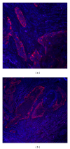
Suppressing endogenous peroxidase is critical for TSA-based in situ hybridization. Residual peroxidase activity from endogenous sources leads to background staining and obscures the truly positive signals. Hydrogen peroxide (H2O2) and hydrochloric acid (HCl) solution were compared for effectiveness in suppression of endogenous peroxidase activity. (a, b) Incubation with high concentration (3%) H2O2 in PBS (instead of more commonly used 0.3% H2O2) for 10 minutes does not suppress endogenous peroxidase. (c, d) The suppression from 0.024 M hydrochloric acid in ethanol for 10 minutes is more extensive. Red: Cy5-tyramide showing existence of peroxidase activity, blue: 4′,6-diamidino-2-phenylindole (DAPI) stained nuclei. Original magnification 200x.
Figure 3
Figure 3
Endogenous biotin in oral cancer tissues. Following satisfactory inhibition of endogenous peroxidase using dilute hydrochloric acid solution (Figure 2), fluorescence in situ hybridization was performed using (a) 5′ biotinylated complementary DNA oligonucleotide probe against miR-146a or (b) control sample treated the same way but without the addition of probe (i.e., without any exogenous biotin). The control sample reveals endogenous biotin in oral cancer tissues. Detection used streptavidin-HRP and Cyanine 5-conjugated tyramide. Red: Cy5-tyramide showing existence of streptavidin-binding activity, blue: DAPI stained nuclei. Original magnification 200x.
Figure 4
Figure 4
miR-146a expression in oral normal squamous epithelia and squamous carcinoma. Microarrayed normal oral tissue (10 cores) and oral squamous cell carcinoma (40 cores) were evaluated using the optimized fluorescence in situ hybridization protocol as outlined in Figure 1. The probe for miR-146a is LNA modified and labeled with digoxigenin at 5′ end. (a, b) Normal oral squamous epithelia. As shown in (a) and (b), cells at the basal layer (adjacent to the white dashed line) are largely negative for miR-146a, while cells at the intermediate layer are positive. In oral squamous carcinomas, (c) well-differentiated tumors, shown here is tissue core B6, often exhibit positive miR-146a staining while (d) poorly differentiated tumors, shown is tissue core E8, tend to be negative. Red: Cy5-tyramide showing positive hybridization signals, blue: DAPI-stained nuclei. Original magnification 400x.

References

    1. Nilsen TW. Mechanisms of microRNA-mediated gene regulation in animal cells. Trends in Genetics. 2007;23(5):243–249. - PubMed
    1. Long D, Lee R, Williams P, Chan CY, Ambros V, Ding Y. Potent effect of target structure on microRNA function. Nature Structural and Molecular Biology. 2007;14(4):287–294. - PubMed
    1. Gaur A, Jewell DA, Liang Y, et al. Characterization of microRNA expression levels and their biological correlates in human cancer cell lines. Cancer Research. 2007;67(6):2456–2468. - PubMed
    1. Zhang L, Yang N, Coukos G. MicroRNA in human cancer: one step forward in diagnosis and treatment. Advances in Experimental Medicine and Biology. 2008;622:69–78. - PubMed
    1. Shen J, Liu N, Todd W, et al. Diagnosis of lung cancer in individuals with solitary pulmonary nodules by plasma microRNA biomarkers. BMC Cancer. 2011;11:p. 374. - PMC - PubMed