. 2012:2012:789232.
doi: 10.1155/2012/789232. Epub 2012 May 10.

Pathogenic Mechanisms and In Vitro Diagnosis of AERD

Affiliations

Pathogenic Mechanisms and In Vitro Diagnosis of AERD

Dirk Schäfer et al. J Allergy (Cairo). 2012.

Abstract

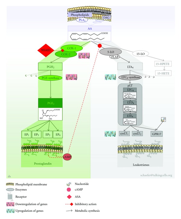
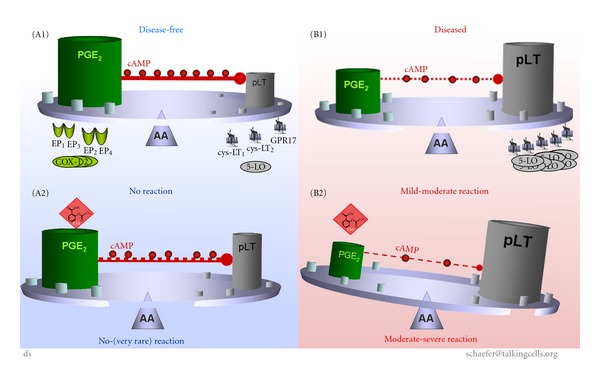
Aspirin-exacerbated respiratory disease (AERD) refers to chronic rhinosinusitis, nasal polyposis, bronchoconstriction, and/or eosinophilic inflammation in asthmatics following the exposure to nonsteroidal anti-inflammatory drugs (NSAIDs). A key pathogenic mechanism associated with AERD is the imbalance of eicosanoid metabolism focusing on prostanoid and leukotriene pathways in airway mucosa as well as blood cells. Genetic and functional metabolic studies on vital and non-vital cells pointed to the variability and the crucial role of lipid mediators in disease susceptibility and their response to medication. Eicosanoids, exemplified by prostaglandin E(2) (PGE(2)) and peptidoleukotrienes (pLT), are potential metabolic biomarkers contributing to the AERD phenotype. Also other mediators are implicated in the progress of AERD. Considering the various pathogenic mechanisms of AERD, a multitude of metabolic and genetic markers is suggested to be implicated and were introduced as potential biomarkers for in vitro diagnosis during the past decades. Deduced from an eicosanoid-related pathogenic mechanism, functional tests balancing PGE(2) and pLT as well as other eicosanoids from preferentially vital leukocytes demonstrated their applicability for in vitro diagnosis of AERD.


Figures

Figure 1
Figure 1
Allocation of terms used for adverse reactions to drugs. The diagram files the term AERD in the context of ADR, drug hypersensitivity, and drug allergy. The terms were gathered from “Report of the Nomenclature Review Committee of the World Allergy Organization” [7], and the proposed classification of allergic and pseudoallergic reactions to drugs that inhibit cyclooxygenase enzymes [9]; AERD: aspirin-exacerbated respiratory diseases, NSAID: nonsteroidal anti-inflammatory drugs, NIUA: NSAID-induced urticaria/angioedema, NBR: NSAID-blended reaction, SDA: single drug-induced anaphylaxis. Definition of ADR according to the World Health Organization [10]: any noxious, unintended, and undesired effect of a drug, which occurs at doses used in humans for prophylaxis, diagnosis, or therapy. This definition excludes therapeutic failures, intentional and accidental poisoning (i.e., overdose), and drug abuse.
Figure 2
Figure 2
COX and 5-LO pathway in the metabolism of PGE2 and pLT for in vitro diagnosis of NSAID-triggered hypersensitivity. Simplified pictogram of eicosanoid pathways in the metabolism PGE2 and leukotrienes implicated for in vitro diagnosis of NSAID-triggered hypersensitivity. AA is enzymatically cleaved by calcium-dependent PLA2 from phospholipids (predominantly) or from DAG (minor amounts). AA is metabolised by the COX-pathway or 5-LO pathway (but also by several other pathways not figured out here). COXs generate PGH2, which is further processed by PGE-synthase forming PGE2 (other PGH2 metabolising pathways not mentioned here). PGE2 binds to EP subtypes of which EP2 and EP4 generate cAMP for signalling cascade. cAMP in turn causes negative feedback on the 5-LO pathway. AA is also metabolised by the 5-LO pathway (in part assisted by FLAP) generating LTA4. LTA4 is further processed by calcium-dependent LTA4-synthase forming amino acids bearing LTC4, which is exported and extracellularly metabolised by enzymes forming LTD4 and LTE4, collectively named pLTs All three LTs bind to cysLTs or GPR17 with differential selectivity. 5-LO: 5-lipoxygenase, AA: arachidonic acid, ASA: acetylsalicylic acid, cAMP: cyclic-adenosine monophosphate; cysLT: receptor of pLT, DAG: diacylglycerole, COX: cyclooxygenase, EP: PGE-receptor,GPR17: orphan receptor, binding pLT and nucleotides, HPETE: hydroxyperoxy-eicosatetraenoic acid, HETE: hydroxy-eicosatetraenoic acid, NSAID: nonsteroidal anti-inflammatory drugs, PLA2: phospholipase A2, PG: prostaglandin, pLT: peptidoleukotrienes.
Figure 3
Figure 3
Causal concept of NSAID-triggered eicosanoid imbalance for in vitro diagnosis of AERD. The causal concept of NSAID-trigger eicosanoid imbalance for in vitro diagnosis of AERD is best allegorised as a tray balancing all parameters (which might be relevant for the pathway) on a needle. Disease-free individuals: housekeeping PGE2 balances synthesis of pLT (e.g., by induction of endogenous cAMP, which inhibits synthesis of pLT); expression of enzymes or receptors are unremarkable (A1). Upon exposure to NSAIDs the PGE2 level is diminished but remains high enough ensuring “uncritical” levels of pLT (even though cAMP might by diminished); expression of enzymes and/or receptors are not modified (A2). Patients with AERD: synthesis of housekeeping PGE2 is diminished, but still balances synthesis of pLT (e.g., by reduced endogenous cAMP); expression of enzymes (up regulation of LTC4-synthase) or receptors (up regulation of cysLT) can be mutated in some cases (B1). Exposure to NSAIDs/aspirin blocks the COX-pathway causing reduced synthesis of PGE2 (and consequently further reduced cAMP level), and consequently the metabolism of arachidonic acid is shifted to the 5LO-pathway provoking elevated synthesis of pLT; expression of enzymes and/or receptors may by altered, but not modified by NSAIDs (B2).
Figure 4
Figure 4
Framework for diagnostic test outcomes. Schema of “real” world diagnostic test outcomes; test measurement: clinical parameters like age, sex, ethnic group, height, weight, and so forth or analytical parameters like temperature, IgE, histamine, interleukins, lipid mediators; shaded areas exemplify the false-positive (false-negative) measurement of disease-free (diseased) individuals, respectively; insert: pictured “ideal” world.
Figure 5
Figure 5
Hypothetical progress of AERD over time.
Figure 6
Figure 6
(a): FET and NSAID-triggered eicosanoid imbalance of individuals suffering from diseases with lower airway symptoms. The FET was performed and the FET values were calculated according to the total eicosanoid pattern score of [11] using PBLs. Patients suffering from NSAID-triggered bronchoconstrictive symptoms were confirmed and characterised by clinical and in vitro diagnosis. Allergy was ruled out by medical history, skin test, and in vitro test for total and specific immunoglobulin. The mean FET value (solid line) of controls, ATA, and AERD was 0.7, 1.4, and 2.1, respectively. FET values > 1.0 characterise patients with lower airway symptoms. FET values ≥ 1, 8 (dashed line, potential threshold) differentiate NSAID-tolerant asthmatics and patients with AERD; ATA: patients suffering from aspirin-tolerant asthma, AERD: patients suffering from aspirin exacerbated respiratory disease; (n = 53 for each group, *P < 0.05, **P < 0.01). (b): FET and functional metabolic differentiation of patients with and without NSAID-triggered eicosanoid of lower and upper airway symptoms. The functional metabolic differentiation (FMD) of subgroups of patient was achieved by in vitro provocation of PBLs and calculation of the FET value according to the total eicosanoid pattern score of [11], but by amending the FET value by subtracting the difference of the sum of the enzymatic capacity (EC) of PG- and LT-synthesis as well as the difference of the ASA- and neuropeptide-induced eicosanoid balances (EB) from the primary FET value (EC and EB were calculated according to [11]). The FET-FMD value takes into account two metabolites of the eicosanoid pathway and their in vitro modification by ASA and neuropeptide. The latter had been shown to be intimately implicated in hyperresponsiveness of airway ([11] and ref. therein). The FET-FMD value reveals the differentiation of ATA, NP, and AERD, but without discrimination of ATA and healthy controls. The mean value of FET-FMD (solid line) was 0.4, 0.4, 1.1, and 1.7, for controls, ATA, NP, and AERD, respectively. The threshold of FET-FMD was ≥1.0 (dashed line) for NSAID-triggered lower and upper symptoms of the airways. In conclusion, this approach confirmed and characterised NSAID-triggered symptoms by clinical and in vitro diagnosis. ATA: patients suffering from bronchial asthma, but tolerant to NSAIDs, NP: patients suffering from nasal polyposis, AERD: patients suffering from aspirin exacerbated respiratory disease with asthmatic symptoms; n = 53 for each group, ns: not significant, *P < 0.05, **P < 0.01. Allergy was ruled out by medical history, skin test and in vitro test of total an specific immunoglobulin.

Similar articles

Cited by

References

    1. Hirschberg. Mitteilungen über einen Fall von Nebenwirkungen des Aspirin. Dt Med Wochenschr. 1902;28:416–417.
    1. Widal F, Abrami P, Lermoyez J. Anaphylaxie et idiosyncrasie. La Presse Medicale. 1922;30:189–193.
    1. Samter M, Beers RF. Concerning the nature of intolerance to aspirin. Journal of Allergy. 1967;40(5):281–293. - PubMed
    1. Delaney JC. The diagnosis of aspirin idiosyncrasy by analgesic challenge. Clinical Allergy. 1976;6(2):177–181. - PubMed
    1. Stevenson DD, Zuraw BL. Pathogenesis of aspirin-exacerbated respiratory disease. Clinical Reviews in Allergy and Immunology. 2003;24(2):169–187. - PubMed

LinkOut - more resources