

doi: 10.3109/10409238.2012.691456.
Epub 2012 Jun 2.
Neuronal regulation of pre-mRNA splicing by polypyrimidine tract binding proteins, PTBP1 and PTBP2
Affiliations
- PMID: 22655688
- PMCID: PMC3422667
- DOI: 10.3109/10409238.2012.691456
Item in Clipboard
Neuronal regulation of pre-mRNA splicing by polypyrimidine tract binding proteins, PTBP1 and PTBP2
Crit Rev Biochem Mol Biol.
2012 Jul-Aug.
Abstract
Alternative splicing patterns are regulated by RNA binding proteins that assemble onto each pre-mRNA to form a complex RNP structure. The polypyrimidine tract binding protein, PTB, has served as an informative model for understanding how RNA binding proteins affect spliceosome assembly and how changes in the expression of these proteins can control complex programs of splicing in tissues. In this review, we describe the mechanisms of splicing regulation by PTB and its function, along with its paralog PTBP2, in neuronal development.
Figures
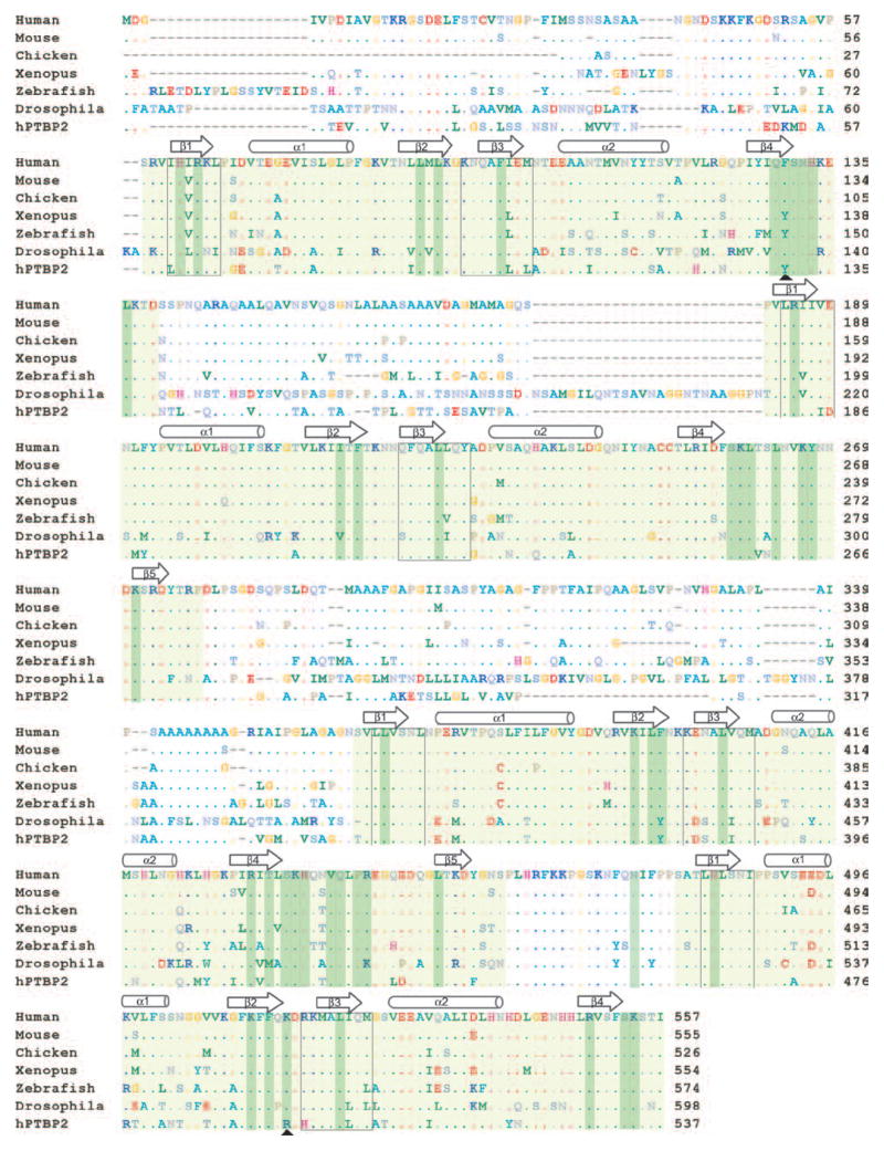
Protein sequence alignment of human, mouse, chicken, Xenopus, Zebrafish, and Drosophila PTB proteins and the human PTBP2 protein. Residues identical to human PTB are shown as dots. RRM domains are shaded light green. RNA interacting residues are shaded dark green. The black boxes indicate the RNP1 and RNP2 motifs. The arrowheads indicate the RNA interacting residues that are different in PTBP2. The N-terminal region of the Drosophila sequence is not shown and the sequence starts at residue 192.
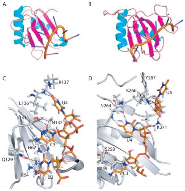
A and B. Ribbon representations of PTB RRMs 1 and 2 bound to a CUCUCU hexamer. The alpha helices are colored cyan, beta strands magenta and loops beige. C and D. Base specific contacts made by RRMs 1 and 2. Amino acids and nucleotides are shown as stick models. The main chain cartoon traces are colored gray. Atomic contacts are indicated by dashed lines.

A. Ribbon representation of PTB RRMs 3 and 4, each bound to a CUCUCU hexamer. B and C. Base specific contacts made by RRMs 3 and 4, respectively. Amino acids and nucleotides are shown as stick models. The main chain cartoon traces are colored gray. Atomic contacts are indicated by dashed lines.
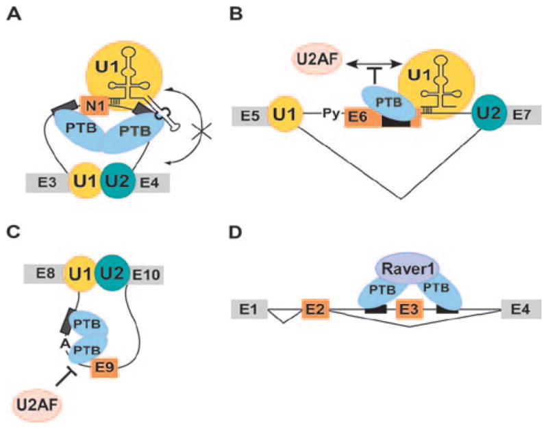
Models for mechanism of splicing repression by PTB. A. PTB binding to introns flanking the c-src N1 exon does not affect 5′ splice site recognition by U1 snRNP but prevents the bound U1 from making intron definition contacts with the components of the 3′ splice site complex at exon 4 downstream. B. In a mechanism similar to c-src N1 exon, binding of PTB to Fas exon 6 does not affect U1 binding. The bound U1 is not able to facilitate formation of the upstream 3′ splice site complex. C. During repression exon E9 of the GABA3 receptor γ2-subunit, PTB binds intronic CU-rich sequences flanking the branchpoint sequence in intron 8 and prevents binding of 3′ splice site factors. D. Repression of tropomyosin exon 3 by PTB requires a cofactor Raver-1, which may bridge the PTB molecules bound to sequences in the flanking introns.
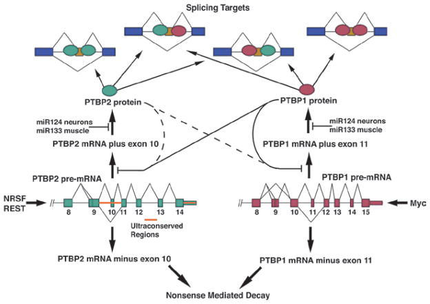
Complex post-transcriptional regulation of PTBP1 and PTBP2. Each gene contains an autoregulated exon whose repression leads to nonsense mediated decay of the transcript. These exons can also be crossregulated by the opposite protein. This is most evident in the repression of PTBP2 exon 10 by PTBP1. Both transcripts are targeted by miRNAs in neurons and muscle cells.

The sequential down regulation of PTB and PTBP2 controls two transitions in splicing programs during neuronal differentiation. Cycling neuronal progenitor cells express PTB but only limited PTBP2. This maintains repression of PTB dependent exons. When PTB is repressed at the onset of differentiation, in part through the action of miR124, PTBP2 is induced. This alters the splicing of transcripts that are more sensitive to PTB than PTBP2 early in neuronal differentiation. PTBP2 expression remains high in differentiating cells. As cells mature and undergo synaptogenesis, PTBP2 expression is reduced. This leads to another splicing regulatory transition, where exons that are effected by both PTB and PTBP2 undergo changes in their splicing.
References
-
- Auweter SD, Oberstrass FC, Allain FH. Solving the structure of PTB in complex with pyrimidine tracts: an NMR study of protein-RNA complexes of weak affinities. J Mol Biol. 2007;367:174–186. - PubMed
Publication types
MeSH terms
Substances
Grants and funding
LinkOut - more resources
Full Text Sources
Other Literature Sources