. 2012 Oct;18(19-20):2073-83.
doi: 10.1089/ten.TEA.2011.0606. Epub 2012 Aug 3.

Anisotropic fibrous scaffolds for articular cartilage regeneration

Affiliations

Anisotropic fibrous scaffolds for articular cartilage regeneration

Seth D McCullen et al. Tissue Eng Part A. 2012 Oct.

Abstract

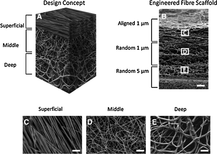
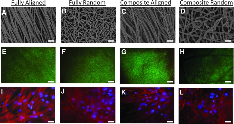
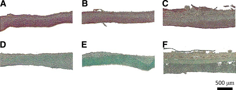
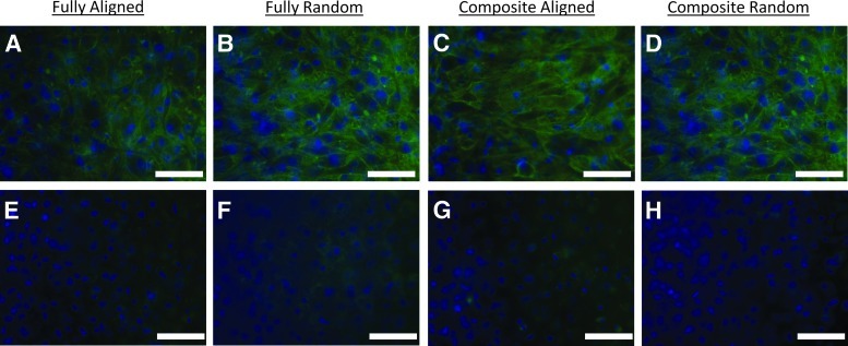
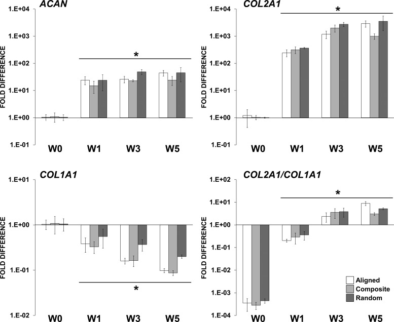
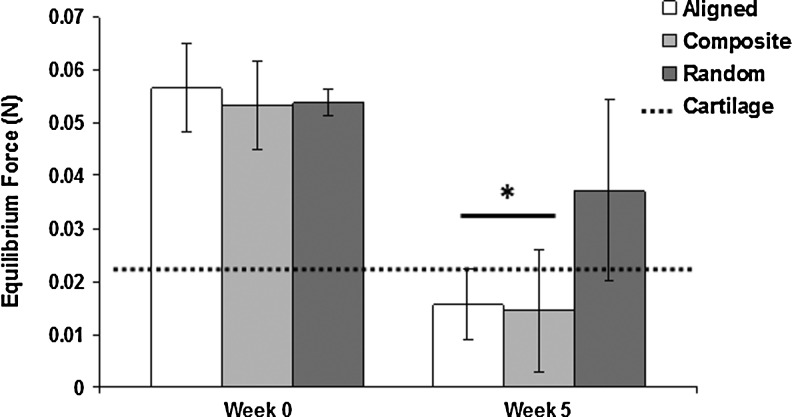
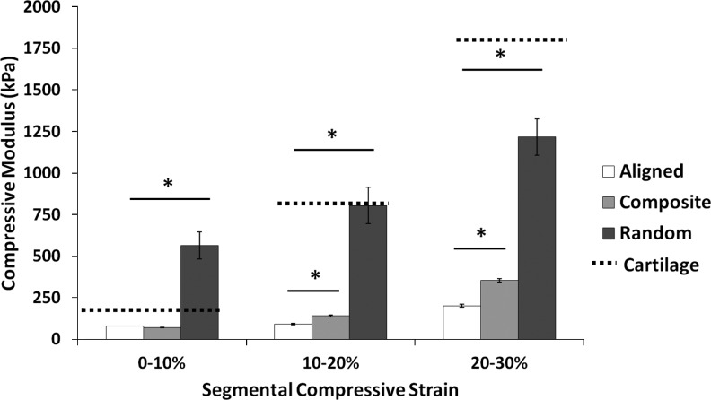
Articular cartilage lesions, which can progress to osteoarthritis, are a particular challenge for regenerative medicine strategies, as cartilage function stems from its complex depth-dependent microstructural organization, mechanical properties, and biochemical composition. Fibrous scaffolds offer a template for cartilage extracellular matrix production; however, the success of homogeneous scaffolds is limited by their inability to mimic the cartilage's zone-specific organization and properties. We fabricated trilaminar scaffolds by sequential electrospinning and varying fiber size and orientation in a continuous construct, to create scaffolds that mimicked the structural organization and mechanical properties of cartilage's collagen fibrillar network. Trilaminar composite scaffolds were then compared to homogeneous aligned or randomly oriented fiber scaffolds to assess in vitro cartilage formation. Bovine chondrocytes proliferated and produced a type II collagen and a sulfated glycosaminoglycan-rich extracellular matrix on all scaffolds. Furthermore, all scaffolds promoted significant upregulation of aggrecan and type II collagen gene expression while downregulating that of type I collagen. Compressive testing at physiological strain levels further demonstrated that the mechanical properties of trilaminar composite scaffolds approached those of native cartilage. Our results demonstrate that trilaminar composite scaffolds mimic key organizational characteristics of native cartilage, support in vitro cartilage formation, and have superior mechanical properties to homogenous scaffolds. We propose that these scaffolds offer promise in regenerative medicine strategies to repair articular cartilage lesions.


Figures

FIG. 1.
FIG. 1.
Sequential electrospinning of poly(ɛ-caprolactone) at varying concentrations and collecting conditions was performed to generate dense fiber networks that preserve zonal fiber mechanical properties in a continuous scaffold. The diagram (A) illustrates the zonal placement of varying fiber zones and the resultant electron microscopy images of the bulk anisotropic scaffold (B) and the varying fibers of aligned 1-μm (C), randomly oriented 1-μm (D), or randomly oriented 5-μm fibers throughout the scaffold (E). Scale bar=100 μm in (B) and 10 μm in (C–E).
FIG. 2.
FIG. 2.
Electron microscopy images of homogeneous fibrous scaffolds of either aligned (A) or randomly oriented fibers (B) and trilaminar scaffolds (C, D) (superficial zone=C and deep cartilage zone=D). Chondrocyte viability on homogenous fibrous scaffolds of either aligned (E) or randomly oriented (F) fiber scaffolds and on trilaminar with either aligned (G) or randomly oriented (H) fiber organizations after five weeks in culture, where live cells fluoresce green and dead cells fluoresce red. Chondrocyte morphology on homogeneous fully fibrous scaffolds of either aligned (I) or randomly oriented (J) fiber scaffolds. Chondrocytes on aligned fibers exhibited spindle-like morphologies orienting along the direction of fibers (I), whereas chondrocytes on randomly oriented scaffolds displayed spread morphologies (J). Chondrocytes on trilaminar scaffolds displayed different morphologies that were dependent on zone-specific fiber orientation and either aligned with fibers (K) or were spread (L). Scale bar=10 μm for (A–D) and (I–L); scale bar=100 μm for (E–H).
FIG. 3.
FIG. 3.
DNA and glycosaminoglycan (GAG) content in chondrocyte-seeded scaffolds during the 5-week culture period. DNA content increased significantly after 1 and 3 weeks in culture, but no further increases were noted at week 5 (A). GAG content significantly increased on week 3 and 5 (B). Normalized GAG content displayed significant enhancements at week 3 and 5 comparative to week 1 (C), indicating significant chondrocyte differentiation. *Significant difference (p<0.05).
FIG. 4.
FIG. 4.
Histological staining of extracellular matrix deposition in fibrous scaffold of aligned (A, B), randomly oriented (C, D), and trilaminar composite scaffolds (E, F) after 5 weeks in culture. Fibrous scaffolds were stained with Picrosirius Red for collagen content (A, C, E) or Alcian Blue for GAG content (B, D, F). Aligned and randomly oriented fibrous scaffolds exhibited homogenous extracellular matrix deposition, whereas in trilaminar composites, matrix deposition was zone specific. Scale bar=500 μm.
FIG. 5.
FIG. 5.
Immunohistochemistry staining for type II collagen (A–D) and type I collagen (E–H) deposition in fibrous scaffold of aligned (A, E), randomly oriented (B, F), and trilaminar composite scaffolds (D, F) after 5 weeks in culture. Blue: nuclei; green: collagen type II (A–D) or collagen type I (E–H); scale bar=50 μm.
FIG. 6.
FIG. 6.
Gene expression profile of bovine chondrocytes cultured on the fibrous scaffolds in a chondrogenic medium. mRNA levels of ACAN, COL2A1, and COL1A1 are represented as fold difference relative to week-0 controls (1 day postseeding) after normalization with the housekeeping GAPDH gene. *Significant difference (p<0.05).
FIG. 7.
FIG. 7.
Equilibrium load attained after a 5-min preload of 0.07N. After five weeks in culture, aligned and trilaminar scaffold groups showed significant decreases in the equilibrium force compared to week-0 controls and were more similar to equilibrium forces measured in native cartilage. *Significant difference (p<0.05).
FIG. 8.
FIG. 8.
Segmental compressive moduli of fibrous scaffolds after 5 weeks in culture. At higher strain rates, significant differences were noted for all scaffold types, and at the higher strain rates, trilaminar composite scaffold exhibited significant increases in compressive moduli compared to aligned fibrous scaffolds. *Significant difference (p<0.05).

References

    1. Daher R.J. Chahine N.O. Greenburg A.S. Sgagilone N.A. Grande D.A. New methods to diagnose and treat cartilage degeneration. Nat Rev Rheumatology. 2009;5:599. - PubMed
    1. Klein T.J. Malda J. Sah R.L. Hutmacher D.W. Tissue engineering of articular cartilage with biomimetic zones. Tissue Eng. 2009;15:143. - PMC - PubMed
    1. Becerra J. Andrades J.A. Guerado E. Zamora-Navas P. Lopez-Puertas J.M. Reddi H. Articular cartilage: structure and regeneration. Tissue Eng Part B. 2010;16:617. - PubMed
    1. Erggelet C. Kreuz P.C. Mrosek E.H. Schagemann J.C. Lahm A. Ducommun P.P., et al. Autologous chondrocyte implantation versus ACI using 3D-bioresorbably graft for the treatment of large full-thickness cartilage lesions of the knee. Arch Orthop Trauma Surg. 2010;130:957. - PubMed
    1. Nuernberger S. Cyran N. Albrecht C. Redl H. Vecsei V. Marlovits S. The influence of scaffold architecture on chondrocyte distribution and behavior in matrix-associated chondrocyte transplantation grafts. Biomaterials. 2011;32:1032. - PubMed

Publication types

LinkOut - more resources