

Longitudinal comparison of diffusion tensor imaging parameters and neuropsychological measures following endoscopic third ventriculostomy for hydrocephalus
- PMID: 22656255
- PMCID: PMC4558885
- DOI: 10.3171/2012.2.PEDS11331
Longitudinal comparison of diffusion tensor imaging parameters and neuropsychological measures following endoscopic third ventriculostomy for hydrocephalus
Abstract
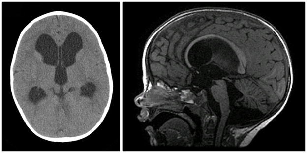
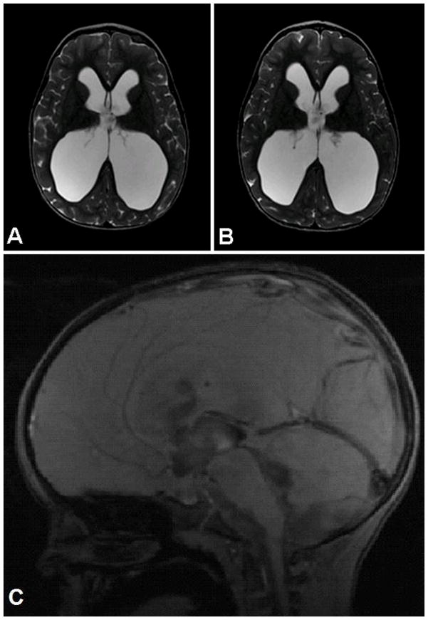
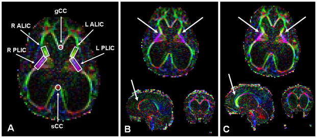
The authors report the case of a 25-month-old boy who underwent endoscopic third ventriculostomy (ETV) for hydrocephalus resulting from aqueductal stenosis. The patient's recovery was monitored longitudinally and prospectively using MR diffusion tensor imaging (DTI) and formal neuropsychological testing. Despite minimal change in ventricle size, improvement in the DTI characteristics and neurodevelopmental trajectory was observed following ETV. These data support the use of DTI as a biomarker to assess therapeutic response in children undergoing surgical treatment for hydrocephalus. In the patient featured in this report, DTI appeared to provide more information regarding postoperative neurodevelopmental outcome than ventricle size alone.
Figures
References
-
- Achenbach TM, Rescorla LA. Manual for the ASEBA Preschool Forms and Profiles. Burlington, VT: University of Vermont Research Center for Children, Youth and Families; 2000.
-
- Air EL, Yuan W, Holland SK, Jones BV, Bierbrauer K, Altaye M, et al. Longitudinal comparison of pre- and postoperative diffusion tensor imaging parameters in young children with hydrocephalus. Clinical article. J Neurosurg Pediatr. 2010;5:385–391. - PubMed
-
- Basser PJ, Pierpaoli C. Microstructural and physiological features of tissues elucidated by quantitative-diffusion-tensor MRI. J Magn Reson B. 1996;111:209–219. - PubMed
-
- Bayley N. Bayley Scales of Infant and Toddler Development. 3. San Antonio, TX: Psychological Corporation; 2005.
