Review
. 2014 Jan 10;20(2):308-24.
doi: 10.1089/ars.2012.4609. Epub 2012 Jul 13.

Mitochondrial redox signaling: Interaction of mitochondrial reactive oxygen species with other sources of oxidative stress

Affiliations
Review

Mitochondrial redox signaling: Interaction of mitochondrial reactive oxygen species with other sources of oxidative stress

Eberhard Schulz et al. Antioxid Redox Signal. .

Abstract

Significance: Oxidative stress is a well established hallmark of cardiovascular disease and there is strong evidence for a causal role of reactive oxygen and nitrogen species (RONS) therein.

Recent advances: Improvement of cardiovascular complications by genetic deletion of RONS producing enzymes and overexpression of RONS degrading enzymes proved the involvement of these species in cardiovascular disease at a molecular level. Vice versa, overexpression of RONS producing enzymes as well as deletion of antioxidant enzymes was demonstrated to aggravate cardiovascular complications.

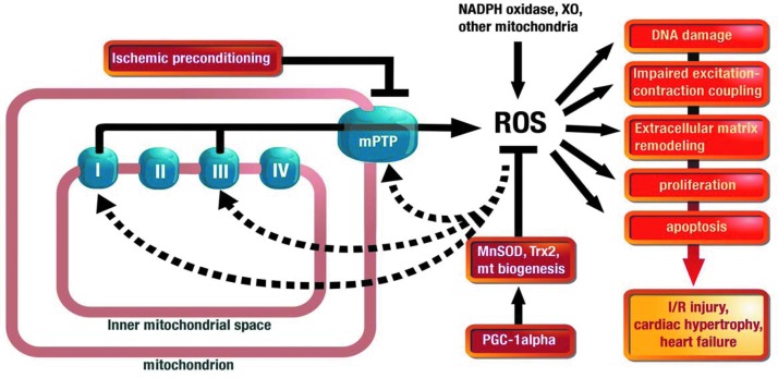
Critical issues: With the present overview we present and discuss different pathways how mitochondrial RONS interact (crosstalk) with other sources of oxidative stress, namely NADPH oxidases, xanthine oxidase and an uncoupled nitric oxide synthase. The potential mechanisms of how this crosstalk proceeds are discussed in detail. Several examples from the literature are summarized (including hypoxia, angiotensin II mediated vascular dysfunction, cellular starvation, nitrate tolerance, aging, hyperglycemia, β-amyloid stress and others) and the underlying mechanisms are put together to a more general concept of redox-based activation of different sources of RONS via enzyme-specific "redox switches". Mitochondria play a key role in this concept providing redox triggers for oxidative damage in the cardiovascular system but also act as amplifiers to increase the burden of oxidative stress.

Future directions: Based on these considerations, the characterization of the role of mitochondrial RONS formation in cardiac disease as well as inflammatory processes but also the role of mitochondria as potential therapeutic targets in these pathophysiological states should be addressed in more detail in the future.


Figures

<b>FIG. 1.</b>
FIG. 1.
Crosstalk between mitochondrial reactive oxygen species (mtROS) and NADPH oxidase–derived ROS. Reverse crosstalk: angiotensin II (AT-II) activates the NADPH oxidase (Nox1 or Nox2) via AT1R-PKC signaling. The resulting superoxide products hydrogen peroxide or peroxynitrite activate the mitochondrial, ATP-sensitive potassium channel (KATP) leading to loss of the mitochondrial membrane potential (mtΨ) with subsequent mtROS formation. mtROS facilitate the opening of the mitochondrial permeability transition pore (mPTP) and subsequent escape of mtROS to the cytosol, where they cause further activation of PKC and NADPH oxidase in concert with increased intracellular calcium. (Classical) crosstalk: Mitochondrial dysfunction [e.g., by nitroglycerin metabolism, MnSOD deficiency, hypoxia, and electron transport chain (ETC) inhibitors] triggers mPTP opening and PKC-NADPH oxidase activation. The resulting vicious circle is suppressed by mtKATP channel blockers (glibenclamide), genetic and pharmacological blockade of the mPTP (CypD−/−, CsA, and SfA), overexpression of MnSOD (MnSODtg) or GPx-1 (GPx-1tg), mitochondria-targeted antioxidants (mitoTEMPO), PKC (Chele), or NADPH oxidase inhibition (p47phox−/−, apocynin) and is aggravated by GPx-1 deficiency (GPx-1−/−). Chele, chelerythrine; CypD−/−, genetic cyclophilin D deletion; MnSODtg, genetic MnSOD overexpression; SfA, sanglifehrin A.
<b>FIG. 2.</b>
FIG. 2.
Redox switches in Nox. Control of translocation: Oxidants from various sources may affect the translocation of important regulatory subunits of Nox2 (and Nox1), p47phox, by oxidation of thiol groups in PKC with subsequent phosphorylation and translocation of these subunits to the membrane causing activation of and superoxide formation by Nox2 (and Nox1). Likewise, those oxidants cause oxidation of thiols in protein disulfide isomerase (PDIox) leading to association with p47phox (maybe also NoxO1 or NoxA1), translocation of this dimer to the membrane, and activation of Nox2 (and Nox1). Reduction of thiols in PDI (PDIred) targets the p47phox complex to the cytosol without activation of Nox. Control of expression: Oxidants contribute to the induction of Nox subunit expression via redox-sensitive transcription factors, but also affect the stability of their mRNA due to post-translational oxidative modification of mRNA-(de)stabilizing proteins, all of which leads to increased expression of Nox subunits. Since Nox4 activity is mainly controlled at the protein level, this pathway may be of great importance for this isoform. The inset shows the phorbol ester/diacylglycerol binding domain of PKCα with two zinc-sulfur clusters (ZnCys3) that function as redox switches in all PKC isoforms. The crystal structure was rendered from the protein database entry 2ELI (DOI:10.2210/pdb2eli/pdb) using the PyMOL Molecular Graphics System Version 1.2r1 (DeLano Scientific LLC). To see this illustration in color, the reader is referred to the web version of this article at www.liebertpub.com/ars
<b>FIG. 3.</b>
FIG. 3.
Redox switches in endothelial nitric oxide synthase (eNOS). X-ray structure of human eNOS with the iron-porphyrin (blue), the substrate L-arginine (green), the P450-forming axial iron-thiolate ligand from a cysteine residue (yellow), the cofactor tetrahydrobiopterin (BH4) (purple), the zinc-thiolate complex forming cysteines (red, two from each subunit), and the zinc ion (brown). The boxes represent the “redox switches” in eNOS, such as S-glutathionylation, PKC- and protein tyrosine kinase-2 (PYK-2)–dependent phosphorylation, oxidative BH4 depletion, disruption of the zinc-sulfur cluster, as well as asymmetric dimethylarginine (ADMA) synthesis/degradation, all of which contribute to the regulation of its enzymatic activity. GSH, glutathione; GSSG, glutathione disulfide. The crystal structure was rendered from the protein database entry 3NOS (DOI:10.2210/pdb3nos/pdb) using the PyMOL Molecular Graphics System Version 1.2r1 (DeLano Scientific LLC). Modified from Daiber and Münzel (26). To see this illustration in color, the reader is referred to the web version of this article at www.liebertpub.com/ars
<b>FIG. 4.</b>
FIG. 4.
Redox switch xanthine dehydrogenase (XDH). Reversible conversion: ROS and reactive oxygen and nitrogen species (RONS) from other sources induce the disulfide formation between cysteine residues 535 and 992, leading to a conformational change of the protein structure with altered affinity for the cofactor NAD+ and subsequent transfer of electrons from the flavin cofactor FADH2 to molecular oxygen. Irreversible conversion: Increased proteolytic activity (ROS and RONS may contribute) causes cleavage and conformational change of the protein structure with altered affinity for the cofactor NAD+ and subsequent transfer of electrons from the flavin cofactor FADH2 to molecular oxygen. FeS, iron-sulfur cluster; MoO2S, molybdo-oxy-thio-complex; RSSR, disulfide bridge. The crystal structure was rendered from the protein database entry 1FIQ (DOI:10.2210/pdb1fiq/pdb) using the PyMOL Molecular Graphics System Version 1.2r1 (DeLano Scientific LLC). To see this illustration in color, the reader is referred to the web version of this article at www.liebertpub.com/ars
<b>FIG. 5.</b>
FIG. 5.
mtROS and cardiac disease. An initial burst of ROS (e.g., from Nox or other mitochondria) leads to the opening of the mPTP and depolarization of the mtΨ. Due to increased electron flux along the ETC, mtROS production will increase (ROS-induced ROS release) and may enter the cytosol via the opened mPTP. mtROS formation can disturb the function of cardiomyocytes in several ways, including DNA damage, impaired excitation-contraction coupling, external matrix remodeling, proliferation, or apoptosis. These molecular changes build the basis for the development of myocardial injury and eventually heart failure. In contrast, ischemic preconditioning prevents myocardial injury by preventing mPTP opening, resulting in decreased mtROS production. Antioxidant enzyme systems represent another strategy to prevent oxidative damage of cardiomyocytes and include MnSOD and thioredoxin-2 (Trx2). Peroxisome proliferator-activated receptor-gamma coactivator-1α (PGC-1α) is an important regulator of this antioxidant defense and may in addition modulate mtROS production by the induction of mitochondrial (mt) biogenesis. To see this illustration in color, the reader is referred to the web version of this article at www.liebertpub.com/ars
<b>FIG. 6.</b>
FIG. 6.
Activation of inflammatory cells by ROS: role of AT-II, mtROS, and other stimuli. Myelomonocytic cells like monocytes contain AT1R and directly respond to AT-II for full activation (2). However, monocytes apparently need to contain the CCR2 [monocyte chemoattractant protein-1 (MCP-1) receptor] (59) and a functional Nox (132) to infiltrate the vascular wall and to induce vascular injury and inflammation in the setting of AT-II hypertension, a process that is paralleled by an increase of proinflammatory chemokines and integrins [i.e., MCP-1 and vascular cellular adhesion molecule-1 (VCAM-1)] in the vascular wall. Interestingly, mtROS that are formed in inflammatory leukocytes dependent on Nox activity, PKC activity, and the uncoupling protein 2 (UCP2) are functionally involved in the setting of AT-II hypertension (37, 42) and atherosclerosis (11, 14). In addition, mitochondrial apoptotic pathways in diabetes that depend on advanced glycation end products (AGE) promote platelet–leukocyte interaction and vascular inflammation (47). The exact nature of the crosstalk of ROS inside inflammatory leukocytes is still a matter of debate.
<b>FIG. 7.</b>
FIG. 7.
Scheme of white blood cell–triggered vascular dysfunction. Infiltrated white blood cells contribute to the development and progression of cardiovascular disease via different pathways. To see this illustration in color, the reader is referred to the web version of this article at www.liebertpub.com/ars

References

    1. Abbas AK. and Lichtman AH. Basic Immunology: Functions and Disorders of the Immune System. Philadelphia, PA: Saunders/Elsevier; 2009, pp. viii, 312
    1. AbdAlla S, Lother H, Langer A, el Faramawy Y, and Quitterer U. Factor XIIIA transglutaminase crosslinks AT1 receptor dimers of monocytes at the onset of atherosclerosis. Cell 119: 343–354, 2004 - PubMed
    1. Abramov AY, Canevari L, and Duchen MR. Beta-amyloid peptides induce mitochondrial dysfunction and oxidative stress in astrocytes and death of neurons through activation of NADPH oxidase. J Neurosci 24: 565–575, 2004 - PMC - PubMed
    1. Ago T, Kuroda J, Pain J, Fu C, Li H, and Sadoshima J. Upregulation of Nox4 by hypertrophic stimuli promotes apoptosis and mitochondrial dysfunction in cardiac myocytes. Circ Res 106: 1253–1264, 2010 - PMC - PubMed
    1. Ago T, Matsushima S, Kuroda J, Zablocki D, Kitazono T, and Sadoshima J. The NADPH oxidase Nox4 and aging in the heart. Aging (Albany NY) 2: 1012–1016, 2010 - PMC - PubMed

Publication types