

Review
doi: 10.1016/j.sbi.2012.04.008.
Epub 2012 May 31.
Packing a punch: the mechanism of pore formation by cholesterol dependent cytolysins and membrane attack complex/perforin-like proteins
Affiliations
- PMID: 22658510
- PMCID: PMC3383384
- DOI: 10.1016/j.sbi.2012.04.008
Item in Clipboard
Review
Packing a punch: the mechanism of pore formation by cholesterol dependent cytolysins and membrane attack complex/perforin-like proteins
Curr Opin Struct Biol.
2012 Jun.
Abstract
The bacterial cholesterol dependent cytolysins (CDCs) and membrane attack complex/perforin-like proteins (MACPF) represent two major branches of a large, exceptionally diverged superfamily. Most characterized CDC/MACPF proteins form large pores that function in immunity, venoms, and pathogenesis. Extensive structural, biochemical and biophysical studies have started to address some of the questions surrounding how the soluble, monomeric form of these remarkable molecules recognize diverse targets and assemble into oligomeric membrane embedded pores. This review explores mechanistic similarities and differences in how CDCs and MACPF proteins form pores.
Copyright © 2012 Elsevier Ltd. All rights reserved.
Figures
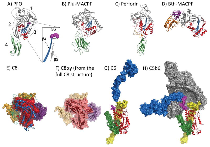
(A) Perfringolysin O (PFO) (PDB ID: 1PFO): the non-contiguous domains are circled and labeled. The CDC/MACPF fold is colored grey with the central β-sheet colored blue and both TMH regions colored red. Domain 4 is a C-terminal immunoglobulin-like fold (green). An inset box shows the β-strands, β4 (blue) and β5 (grey). Magenta spheres show the double glycine motif at the turn of the β-strands. (B) Plu-MACPF (PDB ID: 2QP2): the C-terminal β-prism domain is colored green. CDC/MACPF fold coloured as for A (PFO). (C) Perforin (PDB ID: 3NSJ): the C-terminal EGF-like domain (orange) and C2 domain (green) are highlighted as well as the C-terminal tail (purple) that lies across the surface of the MACPF and EGF-like domains. CDC/MACPF fold coloured as for A (PFO). (D) Bth-MACPF (PDB ID: 3KK7): C-terminal domains include a YegP-like fold (purple) and a second, uncharacterised fold (orange). CDC/MACPF fold coloured as for A (PFO). (E) C8 (PDB ID: 3OJY): The C8α and C8γ components of the C8 structure are shown in surface representation with the MACPF coloured red and the N and C-terminal auxiliary domains coloured yellow. C8γ is coloured purple. The C8β is shown in cartoon representation in front of C8αγ where the MACPF domain is coloured blue and the auxiliary domains are coloured green. (F) The C8αγ component of the whole C8 structure is shown in transparent surface with cartoon representation. Colours as for (E). (G) C6 (PDB ID: 3T5O): The MACPF domain is shown in red cartoon. The N-terminal auxiliary domains are shown as yellow surface. The C-terminal auxiliary domains, EGF and TSP3 domains, are shown in green surface, followed by a segment of the disordered flexible hinge region (magenta surface) that connects to the remaining C-terminal domains (coloured blue). (H) C5b6 (PDB ID: 4A5W): The MACPF domain is shown in red cartoon. The C5b molecule is shown as grey surface representation. The C6 auxiliary domains are colored as for G (C6).
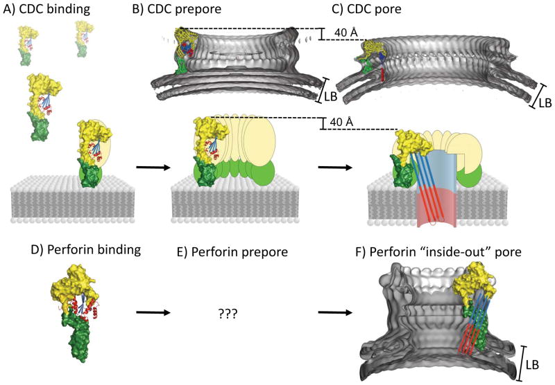
(A) Membrane recognition shows the initial recognition of the lipid bilayer (LB) membrane by domain 4 of a CDC coloured green. The X-ray crystal structure of PFO was used (PDB: 1PFO). Domains 1 and 2 of PFO are coloured yellow. Domain 3 is coloured blue at the β-sheet and red at the α-helices. (B) Oligomerization precedes pore formation in PFO by forming “prepores” [32,33] (C) Pore formation results in channel formed by an extended β-barrel where each molecule contributes 2 β-hairpins (4 β-strands) [25,31] by unraveling the α-helices (red). Shown above panels B and C are the electron density maps created from single particle cryo-electron microscopy of the pneumolysin prepore (EMDB ID: 1106, 28 Å resolution, PDB ID: 2BK2) and pore fitted with the PFO structure (EMDB ID: 1107, 28 Å resolution, PDB ID: 2BK2) [23] (D) The X-ray crystal structure of perforin (PDB: 3NSJ), orientated in the same way as PFO (part A). All C-terminal regions are coloured green, the MACPF domain is coloured yellow and the central β-sheet coloured blue with the bundles of α-helices coloured red. (E) It is unknown whether perforin is able to form a prepore intermediate as seen for CDC’s (F) The electron density map of the mouse perforin pore created from single particle cryo-electron microscopy [7] pore at 28.5 A resolution (EMDB ID: 1769). The putative “inside out” arrangement of the perforin TMHs is shown as an overlaid schematic. LB = Lipid bilayer.
References
-
- Tschopp J, Masson D, Stanley KK. Structural/functional similarity between proteins involved in complement- and cytotoxic T-lymphocyte-mediated cytolysis. Nature. 1986;322:831–834. - PubMed
-
- Nagai H, Oshiro N, Takuwa-Kuroda K, Iwanaga S, Nozaki M, Nakajima T. Novel proteinaceous toxins from the nematocyst venom of the Okinawan sea anemone Phyllodiscus semoni Kwietniewski. Biochem Biophys Res Commun. 2002;294:760–763. - PubMed
-
- Martin JR, Raibaud A, Ollo R. Terminal pattern elements in Drosophila embryo induced by the torso-like protein. Nature. 1994;367:741–745. - PubMed
-
- Zheng C, Heintz N, Hatten ME. CNS gene encoding astrotactin, which supports neuronal migration along glial fibers. Science. 1996;272:417–419. - PubMed
Publication types
MeSH terms
Substances
Grants and funding
LinkOut - more resources
Full Text Sources
Other Literature Sources
Medical
