Review
. 2012 Jun;22(3):304-12.
doi: 10.1016/j.sbi.2012.04.007. Epub 2012 May 31.

Recent developments in single-molecule DNA mechanics

Affiliations
Review

Recent developments in single-molecule DNA mechanics

Zev Bryant et al. Curr Opin Struct Biol. 2012 Jun.

Abstract

Over the past two decades, measurements on individual stretched and twisted DNA molecules have helped define the basic elastic properties of the double helix and enabled real-time functional assays of DNA-associated molecular machines. Recently, new magnetic tweezers approaches for simultaneously measuring freely fluctuating twist and extension have begun to shed light on the structural dynamics of large nucleoprotein complexes. Related technical advances have facilitated direct measurements of DNA torque, contributing to a better understanding of abrupt structural transitions in mechanically stressed DNA. The new measurements have also been exploited in studies that hint at a developing synergistic relationship between single-molecule manipulation and structural DNA nanotechnology.


Figures

Figure 1
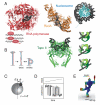
Figure 1. Overview
A) Mechanical processes in DNA biology. RNA polymerase (red, PDB: 1I6H) unwinds DNA during transcript initiation; nucleosomes (blue, PDB: 1AOI), RecA (orange, PDB: 3CMT) distort their substrates. Type II topoisomerases (green, PDB: 2RGR) couple changes in DNA topology to ATP consumption, using a mechanism in which a DNA segment is passed through a transient protein-bridged gap in the duplex. B) Twist (Tw) and writhe (Wr). In micromanipulation experiments, the effective linking number (Lk) of a DNA molecule can be controlled by rotation. Changes in Lk can partition into local Tw and global Wr deformations[55]. C) New methods allow torque measurements in magnetic tweezers, using setups that confine beads to soft angular potentials[21]. D) DNA exhibits rich behavior under torque, such as abrupt buckling transitions[28]. E) Single-molecule measurements of the DNA duplex have been used to parameterize predictions of equilibrium structures and fluctuations in DNA origami [42,45]; heat map indicates predicted RMSD fluctuations.
Figure 2
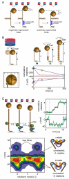
Figure 2. DNA-centric measurements of structural transitions in nucleoprotein complexes
A) Approach based on conventional MT [7,8]. Plectoneme formation provides a large signal reflecting linking number changes trapped in the nucleoprotein complex. Independent contributions to bead height from linking number and DNA contraction can be deconvoluted by repeating measurements on negatively and positively supercoiled DNA. B) Freely Orbiting Magnetic Tweezers (FOMT) assay[10]. A vertically oriented magnetic field allows free rotation of the bead about the DNA axis. In the presence of RecA under 6.5 pN (blue curve) or 1.5 pN (red curve) tension, the bead traces out a spiral as RecA unwinds and lengthens the duplex, reaching saturating values (dashed lines) of angle (linking number) and z expected for complete filament formation on the basis of crystallographic data. C) Rotor bead tracking (RBT) assay. The DNA template is stretched using magnetic tweezers, and a fluorescent rotor bead (diameter ~300 nm) is imaged to obtain angle and z measurements reflecting interactions with protein (blue cylinder). Data are shown from a study of ATP-dependent dynamics in DNA gyrase[11]. Changes in extension and stepwise directional introduction of supercoils can be observed during processive bursts of activity due to individual enzymes. Structural intermediates visited by the enzyme can be mapped out on an angle-extension plane (2D histogram).
Figure 3
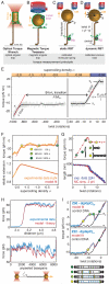
Figure 3. Torque measurements and structural transitions in mechanically stressed DNA
A-D) Methods for measuring torque in single stretched DNA molecules. Tension is applied using force handles (gold) which are often but not always identical with rotational probes (highlighted in green) used to measure torque. A) The optical torque wrench employs a linearly polarized optical trap and nanofabricated birefrigent cylinders[19]. B) In magnetic torque tweezers[21] (MTT) the orientation of the magnetic bead (MB) is measured using marker beads, allowing accurate determination of angular deviations proportional to the applied torque (Fig. 1B). C) In static rotor bead tracking (RBT) the angular position of the rotor bead (RB) may be used to infer the torque based on calibration of the upper transducer DNA segment[26], allowing precise investigation of the response of the lower DNA segment. D) In dynamic RBT, twist introduced by rotating the magnets is relaxed by free rotation of the rotor bead, but a steady-state fixed twist can be maintained using a feedback algorithm[26]. Torque is measured from the RB angular velocity after calibrating its rotational drag. E) Torque-twist diagram of a 4.6 kb DNA, showing characteristic features including the post-buckling torque τB, critical torque for the B-L transition τL, and torsional spring constant formula image (where l is the length of the DNA and Pt,eff is the effective twist persistence length). Δθ0,L, extrapolated change in twist per basepair for the B-L transition; N, number of basepairs of DNA; κL, torsional spring constant for L-DNA. Data are from [26], obtained using dynamic RBT. F) Torque and extension for a complete B-to-L transition measured using an optical torque wrench, and global fit to an analytical model[34]. G) Torque and extension at the plectonemic buckling transition. Extension data are shown together with simulated extension and simulated torque[30]. H) Sequence dependent unpeeling of dsDNA under extension[40], shown together with an equilibrium model based on known basepair stabilities. I) The torsional response of GC repeats[26] is explained by cooperative B-Z transitions. In the depicted microstates, energetically costly junctions between B-form and Z-form are shown as yellow bars.
Figure 4
Figure 4. Mechanics and DNA nanotechnology
A) Snapshots of DNA origami ‘S’-structures during deformation and relaxation in finite element calculations[45] parameterized using single-molecule measurements. In-plane features are reproduced in an averaged TEM image; additional out-of-plane deformations are predictions awaiting confirmation. B) Characterization of DNA origami structures using magnetic tweezers. Oriented rigid attachment of mulithelix bundles is achieved using wide plinth structures with several attachment points. The MB position thermally samples an arc in the xz plane, as expected for oriented attachment of a rigid beam. Torsional persistence lengths of the origami structures can be extracted from slopes in torque-twist plots (right lower panel) and show moderate stiffening in comparison to a single duplex.

Similar articles

Cited by

References

    1. Killian JL, Li M, Sheinin MY, Wang MD. Recent advances in single molecule studies of nucleosomes. Curr Opin Struct Biol. 2012;22(1):80–87. - PMC - PubMed
    1. Aathavan K, Politzer AT, Kaplan A, Moffitt JR, Chemla YR, Grimes S, Jardine PJ, Anderson DL, Bustamante C. Substrate interactions and promiscuity in a viral DNA packaging motor. Nature. 2009;461(7264):669–673. - PMC - PubMed
    1. Bustamante C, Bryant Z, Smith SB. Ten years of tension: Single-molecule DNA mechanics. Nature. 2003;421(6921):423–427. - PubMed
    1. Vijayan V, Zuzow R, O’Shea EK. Oscillations in supercoiling drive circadian gene expression in cyanobacteria. Proc Natl Acad Sci U S A. 2009;106(52):22564–22568. A dramatic demonstration of an organism using mechanical stress in DNA to control complex global changes in transcriptional profiles. The physical basis for differing promoter responses, which are only hinted at by observed sequence biases, deserves further study.

    1. Vologodskii A. Determining protein-induced DNA bending in force-extension experiments: Theoretical analysis. Biophys J. 2009;96(9):3591–3599. - PMC - PubMed

Publication types

LinkOut - more resources