. 2012 Sep;35(9):527-35.
doi: 10.1016/j.tins.2012.04.012. Epub 2012 Jun 2.

Functional network dysfunction in anxiety and anxiety disorders

Affiliations

Functional network dysfunction in anxiety and anxiety disorders

C M Sylvester et al. Trends Neurosci. 2012 Sep.

Abstract

A recent paradigm shift in systems neuroscience is the division of the human brain into functional networks. Functional networks are collections of brain regions with strongly correlated activity both at rest and during cognitive tasks, and each network is believed to implement a different aspect of cognition. We propose here that anxiety disorders and high trait anxiety are associated with a particular pattern of functional network dysfunction: increased functioning of the cingulo-opercular and ventral attention networks as well as decreased functioning of the fronto-parietal and default mode networks. This functional network model can be used to differentiate the pathology of anxiety disorders from other psychiatric illnesses such as major depression and provides targets for novel treatment strategies.


Figures

Figure 1
Figure 1
The human brain can be divided into functional networks. Seven of these networks are illustrated here, including: (a) the four networks discussed in the main text (cingulo-opercular, ventral attention, fronto-parietal, and default mode), and (b) three other commonly described networks (visual, dorsal attention and sensorimotor). Each functional network is believed to implement unique aspects of cognition. This figure is modified, with permission, from a study of the functional network organization of the human brain. Briefly, the investigators measured very low frequency brain activity (<0.08 Hz) using functional magnetic resonance imaging (fMRI) in healthy subjects lying quietly at rest. Correlations in this low frequency brain activity were calculated between all voxel pairs (a voxel is the smallest unit of fMRI data, equivalent to a three dimensional pixel) and each voxel was subsequently categorized into a particular network using graph theory techniques. For visualization purposes, each voxel was color-coded based on network identity and the data were projected onto the cortical surface using CARET software and the Population-Average, Landmark- and Surface-based (PALS) atlas. aDLPFC: anterior dorsolateral PFC; dACC: dorsal anterior cingulate cortex; IPS: intraparietal sulcus; IT: inferior temporal cortex; LP: lateral parietal cortex; MCC: middle cingulate cortex; PCC: posterior cingulate cortex; PCG: pre/post central gyrus; pDLPFC: posterior dorsolateral PFC; PFC: prefrontal cortex; pOcc: posterior occipital cortex; sgACC: subgenual anterior cingulate cortex; SPL: superior parietal lobule; STG: superior temporal gyrus; TPJ: temporal-parietal junction; VLPFC: ventrolateral PFC.
Figure 2
Figure 2
Anxiety disorders may be characterized by a particular pattern of network-level pathology. This Venn diagram highlights changes in behaviors associated with individual networks; anxiety disorders may arise as an interaction between these behaviors. A full description of network-level pathology includes changes in behaviors, task-dependent activity, between-network functional connectivity, and within-network functional connectivity changes associated with each network. Although individuals with all of the network-level changes illustrated above may have the highest probability of developing an anxiety disorder, individuals with most - but not all - of these problematic behaviors may also be at elevated risk. One possibility is that a single network-level alteration triggers a chain of events involving network interactions that eventually results in the full constellation of pathology associated with anxiety disorders. The pathological network-level changes diagrammed above are not meant to be exhaustive. Moreover, there is likely to be heterogeneity even within an individual disorder. Highly co-morbid illnesses such as major depression may arise because of a similar but unique pattern of network-level pathology that is not mutually exclusive with the pattern that characterizes anxiety disorders. Background Venn diagram downloaded from http://en.wikipedia.org/wiki/File:Symmetrical_5-set_Venn_diagram.png on June 11, 2011.
Figure 3
Figure 3
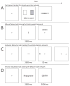
Cognitive training tasks can be designed to correct the network-level pathology of anxiety disorders. A. A task to improve functioning of the cingulo-opercular network during error sensitivity could involve subjects selecting one of two cards on each trial. Error rate is explicitly manipulated by pre-determining on each trial whether the subjects’ response will be correct or incorrect. Starting with a zero error rate and slowly introducing errors, increased cingulo-opercular network activity in response to errors may decrease over many trials through repeated exposure. B. A task to improve functioning of the fronto-parietal network during top-down attentional control could involve repeated practice of a task involving response conflict such as the Erikson flanker task. At the beginning of each trial, a central arrow cues subjects to pay attention to a peripheral location, even as subjects continue to look directly at the central cross. Following a delay, a target appears at the location that had been cued by the central arrow and the subjects’ task is to indicate whether the central letter in the target string is an X or an R. By varying the location of the target letter string as well as the congruency between the central letter and the surrounding distracter letters, subjects may learn through practice to develop improved functioning of the fronto-parietal network and top-down attentional control. C. A task to improve functioning of the ventral attention network during stimulus-driven attention could involve directing subjects to covertly attend to a peripheral location with a central arrow. Following a delay, a dot appears briefly at a task-irrelevant location. On the final frame, subjects determine whether a T at the location cued by the central arrow is upright or inverted. Over many trials, subjects may learn to ignore the task-irrelevant dot, decreasing the influence of stimulus-driven attention. D. A task to improve functioning of the default mode network during emotion regulation could involve directing subjects to use a particular emotion regulation strategy prior to the presentation of a threatening word (task based on ).

References

    1. Lewis CM, et al. Learning sculpts the spontaneous activity of the resting human brain. Proc Natl Acad Sci USA. 2009;106:17558–17563. - PMC - PubMed
    1. Vincent JL, et al. Intrinsic functional architecture in the anaesthetized monkey brain. Nature. 2007;447:83–86. - PubMed
    1. Margulies DS, et al. Precuneus shares intrinsic functional architecture in humans and monkeys. Proc Natl Acad Sci U S A. 2009;106:20069–20074. - PMC - PubMed
    1. Raichle ME. The restless brain. Brain Connectivity. 2011;1:3–12. - PMC - PubMed
    1. Power JD, et al. Functional network organization of the human brain. Neuron. 2011;72:665–678. - PMC - PubMed

Publication types