. 2012 Jun 12;22(6):1299-312.
doi: 10.1016/j.devcel.2012.04.005. Epub 2012 May 31.

Notch ligand endocytosis generates mechanical pulling force dependent on dynamin, epsins, and actin

Affiliations

Notch ligand endocytosis generates mechanical pulling force dependent on dynamin, epsins, and actin

Laurence Meloty-Kapella et al. Dev Cell. .

Abstract

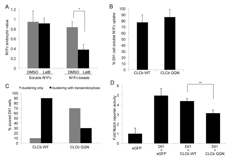
Notch signaling induced by cell surface ligands is critical to development and maintenance of many eukaryotic organisms. Notch and its ligands are integral membrane proteins that facilitate direct cell-cell interactions to activate Notch proteolysis and release the intracellular domain that directs Notch-specific cellular responses. Genetic studies suggest that Notch ligands require endocytosis, ubiquitylation, and epsin endocytic adaptors to activate signaling, but the exact role of ligand endocytosis remains unresolved. Here we characterize a molecularly distinct mode of clathrin-mediated endocytosis requiring ligand ubiquitylation, epsins, and actin for ligand cells to activate signaling in Notch cells. Using a cell-bead optical tweezers system, we obtained evidence for cell-mediated mechanical force dependent on this distinct mode of ligand endocytosis. We propose that the mechanical pulling force produced by endocytosis of Notch-bound ligand drives conformational changes in Notch that permit activating proteolysis.


Figures

Figure 1
Figure 1. Dll1 cells use epsin-dependent CME to activate Notch
(A) Notch reporter activity for HA-N1eGFP expressing cells co-cultured with cells expressing either the endocytic mutant OCDD1 or Dll1 treated with the indicated siRNAs. Values are mean of three independent experiment done in triplicates ± SEM and represent fold-activation over cocultures with parental L cells. *p<0.05 and p**<0.01; Student’s t-test. SCR: scrambled; CHC: clathrin heavy chain; cav-1: caveolin 1. (B) Western blot analysis of Dll1 cells treated with epsin1 and epsin2 siRNAs. α-tubulin indicates equal loading (lower panel). (C-D) Confocal images of coculture assays using HA-N1 cells with (C) Dll1 cells or (D) OCDD1 cells to detect and quantitate NECD transendocytosis. Surface Dll1 (blue), surface HA-N1 (red), post-permeabilizied HA-N1 signal (green) and pre- and post -permeabilization HA-N1 signal overlap (yellow). (E) Quantification of NECD transendocytosis by Dll1 cells treated with indicated siRNAs co-cultured with HA-N1 cells. Values represent the % of Dll1 cells interacting with HA-N1 cells scored for “clustering only” (yellow signals as in D) or “clustering with transendocysosis” (green signals as in C) ± SEM of 3 independent experiments. (F) Representative confocal images used for quantification in Figure 1E. Arrows indicate cell surface HA-N1 clustering; arrowheads indicate internal HA-N1. Bottom images are enlargements of the upper images. (G) Quantification by confocal microscopy of the rescue of NECD transendocytosis defects associated with epsin siRNA knockdown by expression of siRNA-resistant rat epsin1-Venus or rat epsin2-Venus constructs in Dll1 cells co-cultured with HA-N1 cells. Values represents the % of Dll1 cells expressing Venus, epsin1-Venus or epsin2-Venus scored for “clustering with transendocysosis” ± SEM of 3 independent experiments. (see also Figure S1)
Figure 2
Figure 2. Dll1 cell CME of soluble or attached N1Fc require distinct endocytic adaptors
(A) Schematic of staining protocol to detect surface and internal N1Fc signals by FACS analysis to calculate N1Fc endocytic value (see Experimental procedure for details). (B-C) FACS analysis of uptake of (B) soluble N1Fc or (C) N1Fc attached to PrtA-beads by OCDD1, Dll1 or Dll1 cells treated with the indicated siRNAs. Values represent mean of at least 3 independent experiments ± SEM. *p<0.05 and **p<0.01. (D) Quantification by confocal microscopy of soluble preclustered N1Fc uptake by Dll1 cells expressing Venus or dominant-negative epsin1ΔUIM-Venus. Values represent the % Dll1 cells with internal N1Fc signal (n=100) for 3 independent experiments ± SD. (E) Quantification of NECD transendocytosis for Dll1 cells expressing Venus or epsin1ΔUIM-Venus cocultured with HA-N1 cells. Values represent the % of Dll1 cells expressing Venus or epsin1ΔUIM-Venus scored for “clustering only” or “clustering and transendocytosis”. (see also Figure S2)
Figure 3
Figure 3. Distinct requirements for actin polymerization in Dll1 cell CME of attached versus soluble Notch
(A) FACS analysis of soluble N1Fc and N1Fc attached to beads in the presence of LatB. Values represent the mean of 3 independent experiments ± SEM. *p<0.05. (B) Quantification of soluble N1Fc by confocal microscopy of Dll1 cells expressing eGFP-CLCb WT or eGFP-CLCb QQN. Values represent the % Dll1 cells expressing eGFP-CLCb WT or eGFP-CLCb QQN with internal N1Fc signal (n=100) for 3 independent experiments ± SD. (C) Quantification of NECD transendocytosis of Dll1 cells expressing eGFP-CLCb WT or eGFP-CLCb QQN cocultured with HA-N1 cells. Values represent the % of Dll1 cells expressing eGFP-CLCb WT or eGFP-CLCb QQN scored for “clustering only” or “clustering and transendocytosis”. (D) Notch reporter activity for HA-N1eGFP cells co-cultured with L cells transiently expressing eGFP, Dll1+eGFP, Dll1+ eGFP-CLCb WT or Dll1+ eGFP-CLCb QQN. Values are mean of one experiment done in triplicate ± SD p**<0.01. (see also Figure S3)
Figure 4
Figure 4. Notch stimulates Dll1 ubiquitylation and complex formation with epsins
(A) Western blot analysis of lysates from L cells expressing Dll1, HA-Ub and epsin1-Venus incubated on N1Fc- or Fc-coated dishes. Cell lysates were immunoprecipiated (IP) with anti-Dll1 ICD and immunoblotted (IB) with antibodies for HA (top panel), Dll1 and epsin1 (middle panel). Whole cell lysates (WCL) were IB with antibodies for Dll1 and epsin1. Asterisks indicate different molecular weight forms of ubiquitylated Dll1. (B) Short (left panel) and long exposure (right panel) of western blot analysis of lysates from L cells expressing Dll1, HA-Ub, and epsin1-Venus incubated on N1Fc- or Fc-coated dishes and IP with anti-GFP followed by IB with anti-Dll1. (C) Lysates from L cells expressing either Dll1 or epsin1 mixed post-lysis (left lane), or from L cells transfected with both epsin1-Venus and Dll1 incubated on either Fc-(middle lane) or N1Fc-dishes (right lane) IP with anti-GFP followed by IB with anti-Dll1 and anti-epsin. Lower panel: WCLs corresponding to cells in the above panel IB with anti-epsin1 and anti-Dll1. (D) Lysates from L cells transfected with Dll1, HA-Ub and epsin1-Venus incubated with either soluble Fc or N1Fc, or cultured on Fc- or N1Fc-coated dishes were IP with anit-GFP and IB with anti-Dll1 and anti-epsin1. WCLs were IB with anti-Dll1 and anti-epsin1 (lower panels). (E) Lysates from L cells expressing Dll1, HA-Ub and either epsin1-Venus or epsin1ΔUIM-Venus incubated on Fc- or N1Fc-coated dishes were IP with anti-GFP and IB with anti-Dll1 and anti-epsin1. WCLs were IB with anti-Dll1 and anti-epsin1 (lower panels). (F) Lysates from 293T cells transfected with Dll1, HA-Ub, Myc-epsin1, Myc-Mib and increasing amounts of eGFP-PAR-1-T560A were IB with anti-GFP and anti-Myc to detect Mib1 protein. (G) Lysates from 293T cells expressing Dll1, HA-Ub, Myc-epsin1 and increasing amounts of eGFP-PAR-1-T560A were IP with anti-Dll1 and IB with anti-Ub and anti-Dll1 (middle panels) or IP with anti-Myc and IB with anti-epsin1 and anti-Dll1 (bottom panels). WCL were IB with anti-GFP, anti-Myc and anti-Dll1 (top panels). (H) Lysates from 293T expressing Dll1, HA-Ub and either Mib or dominant negative Mib178 were IP with Dll1 and IB with anti-HA, anti-Dll1 and anti-epsin1 (bottom panels). WCL were IB with anti-Myc and anti-Dll1 (top panels). (I) Lysates from L cells expressing Dll1, HA-Ub and epsin1-Venus treated with Mib1 or SCR siRNAs were IP with anti-Dll1 and IB with anti-Mib1 and anti-Dll1 (middle panels) or IP with anti-GFP and IB with anti-epsin and anti-Dll1 (bottom panels). WCL were IB with anti-Mib1, anti-Dll1 and anti-epsin1 (top panels).
Figure 5
Figure 5. Laser tweezers detect mechanical forces exerted by ligand cells on trapped N1Fc-beads
(A) Schematic of optical tweezers system used to measure Dll1 cell-mediated forces exerted on trapped N1Fc-beads. (B) Prototypic force tracing for Dll1 cells bound to laser trapped N1Fc-beads. (C) Prototypic force tracing for Dll1 cells interacting with uncoated PrtA-beads. (D) Prototypic force tracing for Dll1 cells interacting with Fc-beads. See also Figure S4 (E) Average of the average force measurement for Dll1 cells interacting with PrtA-, Fc- or N1Fc-beads. **p< 0.01; ***p <0.001. see also Table1 (F-G) Prototypic force tracings for OCDD1 cells interacting with (F) N1Fc- or (G) Fc-beads. (H) Average of the average force measurement for OCDD1 cells interacting with Fc- or N1Fc-beads. *p < 0.05. see also Table1 (I) Average of the average force measurement for Dll1 cells and OCDD1 cells interacting with N1Fc-beads. ***p <0.001. see also Table1
Figure 6
Figure 6. Dll1 cells pull on laser trapped N1Fc beads with sustained force requiring endocytosis dependent on dynamin, epsins and actin
(A-B, D-E, G-H, J-K, M-N) Prototypic force tracings for Dll1 cells expressing (A) eGFP or, (B) DynK44A-eGFP, (G) epsin1ΔUIM-Venus or (H) Venus, (J) eGFP-PAR-1 or (K) eGFP-PAR-1T560A, (M) CLCb QQN-eGFP or (N) CLCb WT-eGFP or (D) treated with DMSO or (E) Dynasore when bound to trapped N1Fc-beads. (C, F, I, L, O) Average of the average force measurement for Dll1 cells expressing (C) eGFP, DynK44A-eGFP or Rab11S25N-eGFP, (I) Venus or epsin1ΔUIM-Venus, (L) eGFP, eGFP-PAR-1 or eGFP-PAR-1T560A, (O) CLCb QQN-eGFP or CLCb WT-eGFP or (F) for Dll1 cells untreated or treated with DMSO or Dynasore. *p < 0.05, **p< 0.01, ***p <0.001. see also Table1

References

    1. Aghamohammadzadeh S, Ayscough KR. Differential requirements for actin during yeast and mammalian endocytosis. Nat Cell Biol. 2009;11:1039–1042. - PMC - PubMed
    1. Aguilar RC, Longhi SA, Shaw JD, Yeh LY, Kim S, Schon A, Freire E, Hsu A, McCormick WK, Watson HA, et al. Epsin N-terminal homology domains perform an essential function regulating Cdc42 through binding Cdc42 GTPase-activating proteins. Proc Natl Acad Sci U S A. 2006;103:4116–4121. - PMC - PubMed
    1. Banks SM, Cho B, Eun SH, Lee JH, Windler SL, Xie X, Bilder D, Fischer JA. The functions of auxilin and Rab11 in Drosophila suggest that the fundamental role of ligand endocytosis in notch signaling cells is not recycling. PLoS ONE. 2011;6:e18259. - PMC - PubMed
    1. Bozkulak EC, Weinmaster G. Selective use of ADAM10 and ADAM17 in activation of Notch1 signaling. Mol Cell Biol. 2009;29:5679–5695. - PMC - PubMed
    1. Brady RJ, Damer CK, Heuser JE, O’Halloran TJ. Regulation of Hip1r by epsin controls the temporal and spatial coupling of actin filaments to clathrin-coated pits. J Cell Sci. 2010;123:3652–3661. - PMC - PubMed

Publication types