. 2012 Aug 15;427(2):164-74.
doi: 10.1016/j.ab.2012.05.019. Epub 2012 May 29.

Fluorescence polarization assay for inhibitors of the kinase domain of receptor interacting protein 1

Affiliations

Fluorescence polarization assay for inhibitors of the kinase domain of receptor interacting protein 1

Jenny L Maki et al. Anal Biochem. .

Abstract

Necrotic cell death is prevalent in many different pathological disease states and in traumatic injury. Necroptosis is a form of necrosis that stems from specific signaling pathways, with the key regulator being receptor interacting protein 1 (RIP1), a serine/threonine kinase. Specific inhibitors of RIP1, termed necrostatins, are potent inhibitors of necroptosis. Necrostatins are structurally distinct from one another yet still possess the ability to inhibit RIP1 kinase activity. To further understand the differences in the binding of the various necrostatins to RIP1 and to develop a robust high-throughput screening (HTS) assay, which can be used to identify new classes of RIP1 inhibitors, we synthesized fluorescein derivatives of Necrostatin-1 (Nec-1) and Nec-3. These compounds were used to establish a fluorescence polarization (FP) assay to directly measure the binding of necrostatins to RIP1 kinase. The fluorescein-labeled compounds are well suited for HTS because the assays have a dimethyl sulfoxide (DMSO) tolerance up to 5% and Z' scores of 0.62 (fluorescein-Nec-1) and 0.57 (fluorescein-Nec-3). In addition, results obtained from the FP assays and ligand docking studies provide insights into the putative binding sites of Nec-1, Nec-3, and Nec-4.


Figures

Figure 1
Figure 1
(A) Structures of the different necrostatins. (B) Induced fit docking model of Nec-1-RIP1. Nec-1 (3) was docked into the molecular model of RIP1 kinase domain (aa 22–291) as described in the Materials and Methods. RIP1 is shown as a cartoon with the N-terminal domain in cyan, P-loop in blue, hinge in magenta, DLG in black, activation segment in orange, and C-terminal domain in green with 3 shown as sticks in red. (C) A surface representation zoomed in to the ATP-binding region of RIP1 is shown with Nec-1 and fluorescein portions of 20 labeled. Different views of this complex are shown in Figure S1. The model of RIP1/20 complex indicates that fluorescein does not interact with RIP1. RIP1 is color-coded as described in B and 20 is color-coded by element: carbon in yellow, nitrogen in blue, and oxygen in red. Images were made using PyMol.
Figure 2
Figure 2
(A) Synthesis of Nec-1 derivatives 4, 5, and 6. Compound 10 was made as previously described [15]. a) various benzyl bromide, KOH, EtOH/H2O, 95°C, 14h. (B) The dose-response curves of optimized racemic Nec-1 (Rac-3), intermediate analogs (4, 5, 6), and Nec-3 (7) in a necroptosis cell-based assay as previously described [14] [15]. Viability was normalized to cells treated with DMSO. (C) In vitro kinase assay of Nec-1 intermediate analogs. The autophosphorylation of recombinant GST-RIP1 was performed as indicated in the Materials and Methods. Samples were run on SDS-PAGE gel and autophosphorylation was visualized by autoradiography. D is the DMSO control sample while the concentrations of Nec-1 analogs are shown above the gel. (D) In vitro kinase assay with the Rac-3, 20, and inactive Nec-1 (2). Compound 20 is able to inhibit the autophosphorylation activity of GST-RIP1. (E) Synthesis of Nec-1 fluorescein derivative. a) triflic anhydride, Py, CH2Cl2, 0°C, 6h; b) Zn(CN)2, Pd(PPh3)4, DMF, 60°C, 14h; c) NBS, AIBN, CCl4, reflux, 12h; d) N,N-dimethylmethylene ammonium chloride, CH2Cl2, rt, 2h, then 1M NaOH; e) NaCN, EtOAc, DMSO, 80°C, 6h; f) DIBAL, toluene, −78°C to rt, 2h; g) (NH4)2CO3, KCN, EtOH, H2O, 60°C, 14h; h) 15, KOH, EtOH/H2O, 95°C, 8h; i) H2, Raney Ni, NH3 in MeOH, 6h; j) fluorescein isothiocyanate (FITC), DMF, 0°C to rt, 1h.
Figure 3
Figure 3
(A) Synthesis of Nec-3 fluorescein derivative. a) NaOH, EtOH, H2O, rt, 2h; b) (CH3)3CCOOH, hydrazine hydrate, EtOH, 120°C, reflux, 16h; c) Methyl 10-chloro-10-oxodecanoate, sat. aq. NaHCO3, EtOAc, 2h; d) NaOH, MeOH, 6h; e) N-Boc-piperazine, HBTU, DIPEA, CH2Cl2, rt, 14h; f) TFA, CH2Cl2, rt, 1h; g) FITC, DMF, 0°C to rt, 1h. (B) In vitro kinase assay with the Nec-3 (7), fluorescein-Nec-3 (26), and inactive Nec-3 (8). Compound 26 is able to inhibit the autophosphorylation activity of GST-RIP1.
Figure 4
Figure 4
KD determination of fluorescent analogs. (A) Increasing concentrations of GST-RIP1 protein were titrated into 20 and the curve was fitted to obtain a KD of 2.6 ± 0.3 µM with a R2 value of 0.96 and Bmax value of 210 mP. (B) Increasing concentrations of GST-RIP1 protein were titrated into 26 and the curve was fitted to obtain a KD of 3.4 ± 0.5 µM with a R2 value of 0.96 and Bmax value of 188 mP.
Figure 5
Figure 5
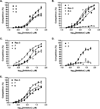
Competition Assays with fluorescent analogs. (A) GST-RIP1 and 20 were incubated together for five minutes and then increasing concentrations of 4, 5, and 6 were titrated. Curves were fit to determine IC50 and Ki values for each compound (Table 1 and 2). (B) Competition assays with GST-RIP1/20 and 1, 2, Rac-3, and R-3. (C) Competition assays with GST-RIP1/20 and Rac-3, 7, and 9. (D) Competition assays with GST-RIP1/26 and 7 and 8. (E) Competition assays with GST-RIP1/26 and Rac-3, 7, and 9.
Figure 6
Figure 6
Docking of the necrostatins to RIP1. (A) The induced fit docking model of Nec-1, color-coded as in Figure 1C, and RIP1 is shown. The residues making the binding pocket for Nec-1 determined from ligand interaction diagram (Figure S2) are color-coded by amino acid type: green-hydrophobic, cyan-polar, red-acidic, blue-basic, and glycinetan. The black dashed line indicates a hydrogen bond. (B) Nec-1 (3) in red and Nec-3 (7) in yellow are docked into the DLG out conformation of RIP1. The two molecules partially overlap in their binding sites. (C) Nec-1 (3) in red and Nec-4 (9) in brown docked into the DLG out conformation of RIP1. The two molecules partially overlap. (D) Nec-3 (7) in yellow and Nec-4 (9) in brown are docked into the DLG out conformation of RIP1. The two molecules significantly overlap with each other.
Figure 7
Figure 7
Feasibility of FP assay for HTS. (A) DMSO tolerance of 20 in FP was tested with 20 alone (black bars) and GST-RIP1/20 complex (white bars) in 0–10% DMSO. Concentrations at or below 5% are well-tolerated in this assay. (B) Z'-score for 20 was tested in three separate experiments in 10-wells each with GST-RIP1/20 and GST-RIP1/20/Rac-3. The average from all 30 wells was used to normalize each of the three separate experiments. Z' is 0.62 demonstrating that this is an excellent assay for HTS. (C) Same as in (A) except using 26. (D) Z'-score for 26 was tested as described in (B) with GST-RIP1/26 and GST-RIP1/26/7. Z' is 0.57 demonstrating that this is an excellent assay for HTS.

References

    1. Dunai Z, Bauer PI, Mihalik R. Necroptosis: Biochemical, Physiological and Pathological Aspects. Pathol. Oncol. Res. 2011 - PubMed
    1. Galluzzi L, Vanden Berghe T, Vanlangenakker N, Buettner S, Eisenberg T, Vandenabeele P, Madeo F, Kroemer G. Programmed necrosis from molecules to health and disease. Int. Rev. Cell Mol. Biol. 2011;289:1–35. - PubMed
    1. Cho YS, Challa S, Moquin D, Genga R, Ray TD, Guildford M, Chan FK. Phosphorylation-driven assembly of the RIP1-RIP3 complex regulates programmed necrosis and virus-induced inflammation. Cell. 2009;137:1112–1123. - PMC - PubMed
    1. Zhang DW, Shao J, Lin J, Zhang N, Lu BJ, Lin SC, Dong MQ, Han J. RIP3, an energy metabolism regulator that switches TNF-induced cell death from apoptosis to necrosis. Science. 2009;325:332–336. - PubMed
    1. He S, Wang L, Miao L, Wang T, Du F, Zhao L, Wang X. Receptor interacting protein kinase-3 determines cellular necrotic response to TNF-alpha. Cell. 2009;137:1100–1111. - PubMed

Publication types

MeSH terms