. 2012 Jul;144(1):1-12.
doi: 10.1016/j.clim.2012.04.001. Epub 2012 Apr 20.

Estrogen receptor alpha modulates Toll-like receptor signaling in murine lupus

Affiliations

Estrogen receptor alpha modulates Toll-like receptor signaling in murine lupus

Melissa A Cunningham et al. Clin Immunol. 2012 Jul.

Abstract

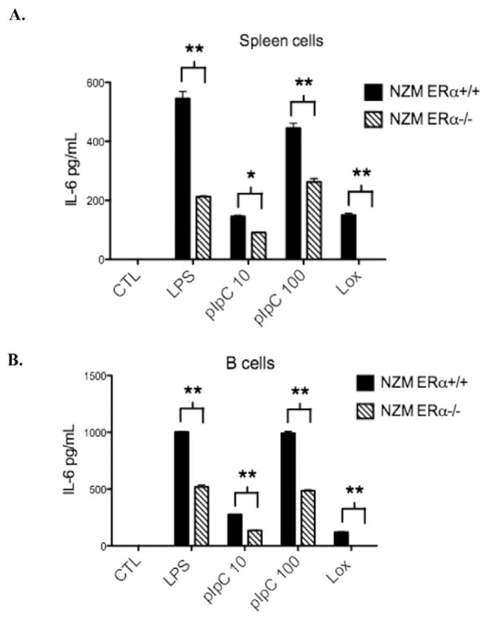
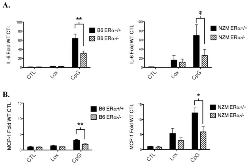
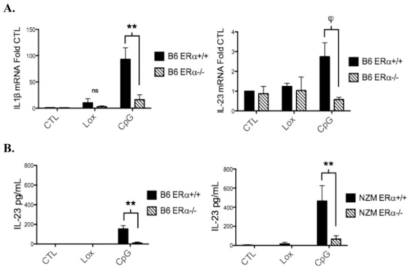
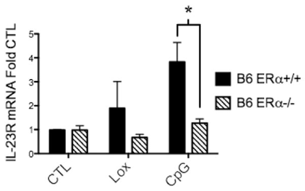
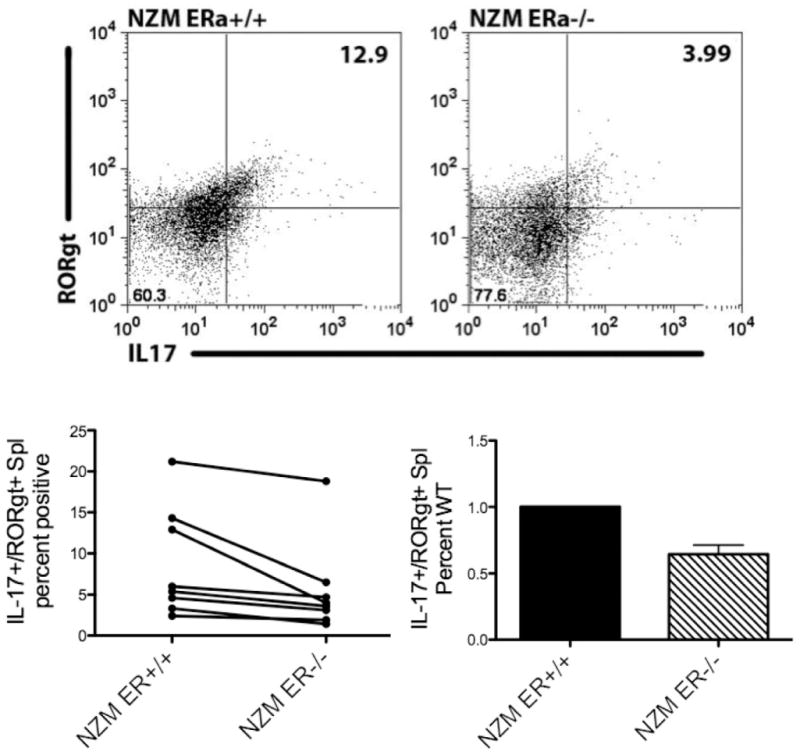
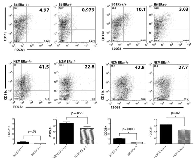
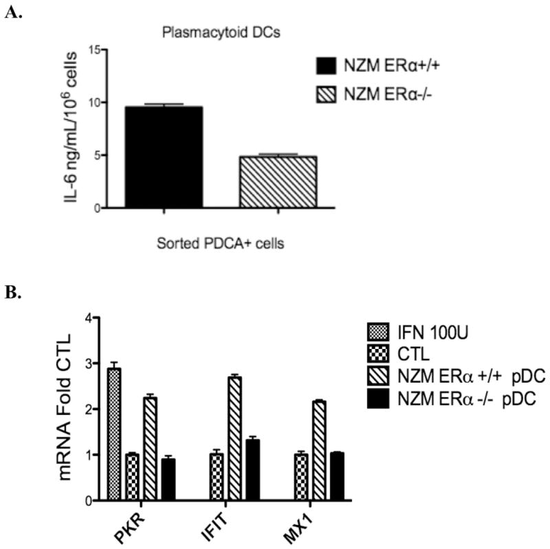
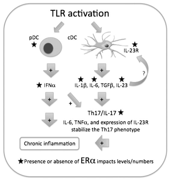
Systemic lupus erythematosus (SLE) is a disease that disproportionately affects females. Despite significant research effort, the mechanisms underlying the female predominance in this disease are largely unknown. Previously, we showed that estrogen receptor alpha knockout (ERαKO) lupus prone female mice had significantly less pathologic renal disease and proteinuria, and significantly prolonged survival. Since autoantibody levels and number and percentage of B/T cells were not significantly impacted by ERα genotype, we hypothesized that the primary benefit of ERα deficiency in lupus nephritis was via modulation of the innate immune response. Using BMDCs and spleen cells/B cells from female wild-type or ERαKO mice, we found that ERαKO-derived cells have a significantly reduced inflammatory response after stimulation with TLR agonists. Our results indicate that the inflammatory response to TLR ligands is significantly impacted by the presence of ERα despite the absence of estradiol, and may partially explain the protective effect of ERα deficiency in lupus-prone animals.


Conflict of interest statement

Conflict of interest statement

The authors declare that there are no conflicts of interest.

Figures

Figure 1
Figure 1
TLR-induced IL-6 production is significantly reduced in isolated spleen cells (A) and B cells (B) from ERα−/− mice. Treatment with a TLR4 agonist (LPS 1ug/ml), TLR3 agonist (polyI:C 10 or 100ug/ml) or a TLR7 agonist (loxoribine 200 mmol) for 24 h resulted in IL-6 induction that was reduced by at least 50% in spleen cells or B cells from ERα−/− animals. Representative of 3 independent experiments with a total of 10 animals per group minimum. *=p<0.05, **=p<0.001.
Figure 2
Figure 2
TLR9-induced IL-6 and MCP-1 production by bone-marrow derived dendritic cells cultured from ERα−/− mice is reduced in both B6 and NZM2410 strains. BMDCs from ERα+/+ and ERα−/− mice were cultured for 24 h with vehicle (CTL), loxoribine (Lox) 50 nmol, or CpG 1ug/ml under estradiol-free conditions. Media was removed and IL-6 (A) or MCP-1 (B) was measured by ELISA. Cytokine levels were in the range of 120 pg/ml–47 ng/ml. Pooled data from n=4 separate experiments with total number of 12 animals per group minimum. ϕ=p<.05, *=p<.01, **=p<0.0001.
Figure 3
Figure 3
TLR9-induced mRNA expression of the inflammatory cytokines IL-1β and IL-23 by dendritic cells is reduced in B6 ERα−/−mice. BMDCs from ERα+/+ and ERα−/− mice were cultured for 24 h with vehicle (CTL), loxoribine (Lox) 50 nmol, or CpG 1ug/ml under estradiol-free conditions. A, RNA was isolated and IL-1β and IL-23 mRNA were measured by qRT-PCR. B, IL-23 protein levels were measured by ELISA (p19-specific capture antibody and a p40 specific detection antibody). Cytokine levels were in the range of 60 pg/ml–700 pg/ml. CpG-induced IL-23 production was reduced in ERα−/− animals in both B6 and NZM strains, n=3 or 4 separate experiments, **=p<0.001, ϕ=p<.05.
Figure 4
Figure 4
TLR9-induced mRNA expression of IL-23R by dendritic cells was reduced in B6 ERα−/− mice. BMDCs from ERα+/+ and ERα−/− mice were cultured for 24 h with vehicle (CTL), loxoribine (Lox) 50 nmol, or CpG 1ug/ml under estradiol-free conditions. RNA was isolated from DCs and IL-23R was measured by qRT-PCR. A similar trend was observed in NZM and MRL/lpr mice that reached statistical significance in MRL DCs and spleen cells (data not shown), n=3 separate experiments, min. 5 animals per group, *=p<0.01.
Figure 5
Figure 5
Unstimulated spleen cells derived from NZM ERα−/− animals displayed reduced numbers of IL-17+/RORγt+cells. Spleen cells were isolated from NZM ERα+/+ and NZM ERα−/− mice and cultured for 24 h +/− GolgiStop. Cells were fixed, permeabilized and stained with IL-17-PE and RORγt-APC. Isotype controls: rat IgG2a for IL-17 and anti-human/anti-mouse IgG mix for RORγt were used to set gates. Results are compiled from 8 sets of animals, n=5 separate experiments. For the difference between WT and ERαKO, both a paired T test and a sign test indicate a significantly larger percent of IL17+/RORγt+spleen cells in WT relative to KO animals (p=0.025 paired t-test, p=0.008 sign test).
Figure 6
Figure 6
BMDCs from lupus-prone mice (middle panel) display increased plasmacytoid DC markers compared with wild-type B6 mice (top panel), despite culturing under traditional cDC conditions, using 2 different pDC-specific markers (PDCA or 120 G8). There is a clear trend toward reduced numbers of PDCA+or 120 G8+ cells in ERα−/− BMDCs which is statistically significant in B6 animals and in NZM mice staining with 120 G8. Flow cytometry plots represent 3 separate experiments, n=6 B6 and n=15 NZM animals (compiled in bottom panel).
Figure 7
Figure 7
IL-6 (A) and IFNα (B) production by plasmacytoid dendritic cells derived from ERα−/− lupus-prone mice is significantly reduced. The number of PDCA+cells after cell-sorting was standardized to 1.0×10⁶ cells/ml and cells were treated for 24 h with CpG 1ug/ml. A, IL-6 production was measured by ELISA and was significantly diminished in ERα−/− pDCs taken from NZM2410 mice, p<.001, representative of 2 independent experiments. B, IFNα activity was measured via WISH assay using conditioned media taken from CpG-treated pDCs. WISH cells were incubated with control media +/− Type I Interferon 100 units or with conditioned media from ERα+/+ or ERα−/− pDCs for 6 h. qRT-PCR was done using IFNα-responsive gene primer sets. ERα+/+ vs. ERα−/− are significantly different, p=.003, representative of 2 independent experiments.
Figure 8
Figure 8
Working model for how ERα may modulate signaling by TLR-activated DCs. Activated DCs produce pro-inflammatory cytokines (ex. IL-23, IL-1β, IFNα) that promote ongoing inflammation via multiple mechanisms. As demonstrated, the expansion and stabilization of the Th17 subset of T cells via IL-6 and IL-23 and the upregulation of IL23R are all impacted by the presence of ERα. In the absence of ERα, TLR stimulation cannot fully induce IL-6, IL-1β, IL-23 or IL-23R upregulation on spleen cells (or DCs), leading to fewer IL-17-producing cells. Additionally, lupus-prone ERα−/−mice have decreased numbers of plasmacytoid DCs that feed into this pathway via production of IFNα.

Comment in

  • Why do women get lupus?
    Moulton VR, Tsokos GC. Moulton VR, et al. Clin Immunol. 2012 Jul;144(1):53-6. doi: 10.1016/j.clim.2012.04.003. Epub 2012 Apr 27. Clin Immunol. 2012. PMID: 22659035 No abstract available.

References

    1. Cunningham M, Gilkeson G. Estrogen receptors in immunity and autoimmunity. Clin Rev Allergy Immunol. 2011;40:66–73. - PubMed
    1. Biswas DK, Singh S, Shi Q, Pardee AB, Iglehart JD. Crossroads of estrogen receptor and NF-kappaB signaling. Sci STKE. 2005;2005:pe27. - PubMed
    1. Jakacka M, Ito M, Weiss J, Chien PY, Gehm BD, Jameson JL. Estrogen receptor binding to DNA is not required for its activity through the nonclassical AP1 pathway. J Biol Chem. 2001;276:13615–13621. - PubMed
    1. Safe S. Transcriptional activation of genes by 17 beta-estradiol through estrogen receptor-Sp1 interactions. Vitam Horm. 2001;62:231–252. - PubMed
    1. Lambert KC, Curran EM, Judy BM, Lubahn DB, Estes DM. Estrogen receptor-alpha deficiency promotes increased TNF-alpha secretion and bacterial killing by murine macrophages in response to microbial stimuli in vitro. J Leukoc Biol. 2004;75:1166–1172. - PubMed

MeSH terms

LinkOut - more resources