. 2013 Mar:66:302-10.
doi: 10.1016/j.neuropharm.2012.05.038. Epub 2012 May 30.

The interaction between mGluR1 and the calcium channel Cav₂.₁ preserves coupling in the presence of long Homer proteins

Affiliations

The interaction between mGluR1 and the calcium channel Cav₂.₁ preserves coupling in the presence of long Homer proteins

Donald Beqollari et al. Neuropharmacology. 2013 Mar.

Abstract

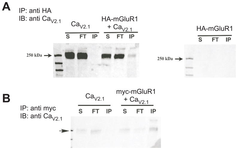
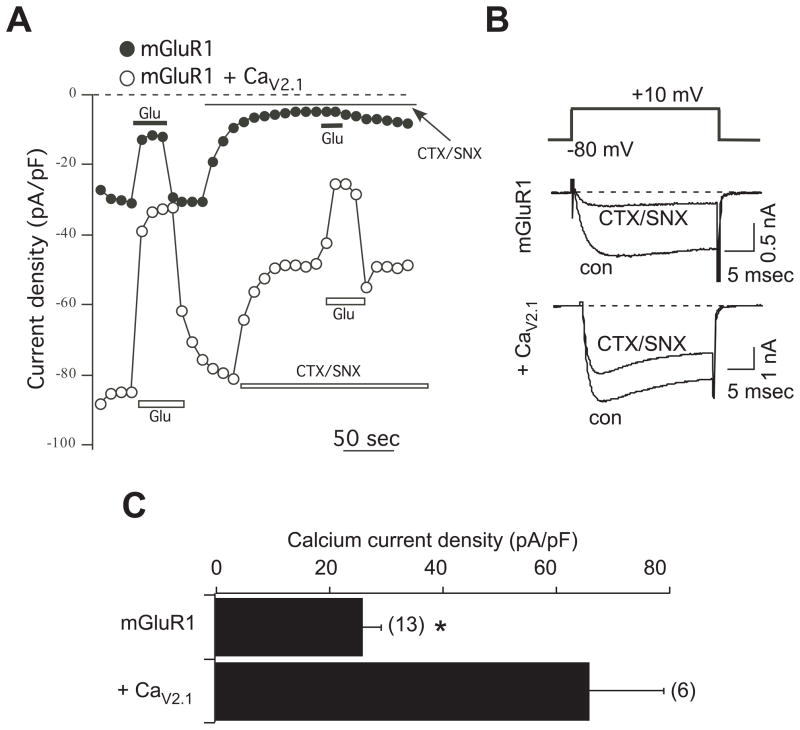
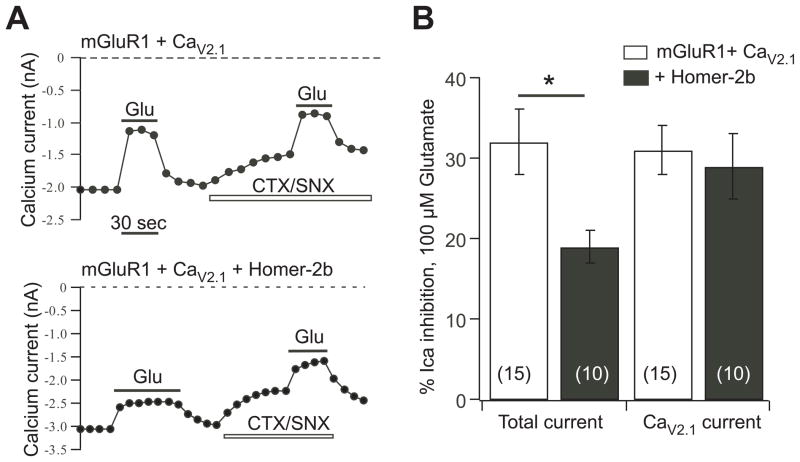
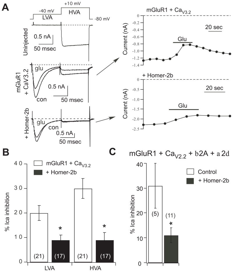
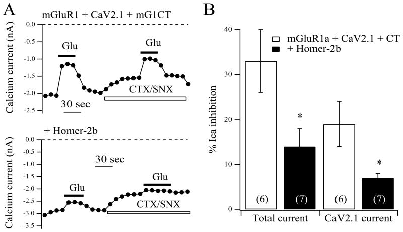
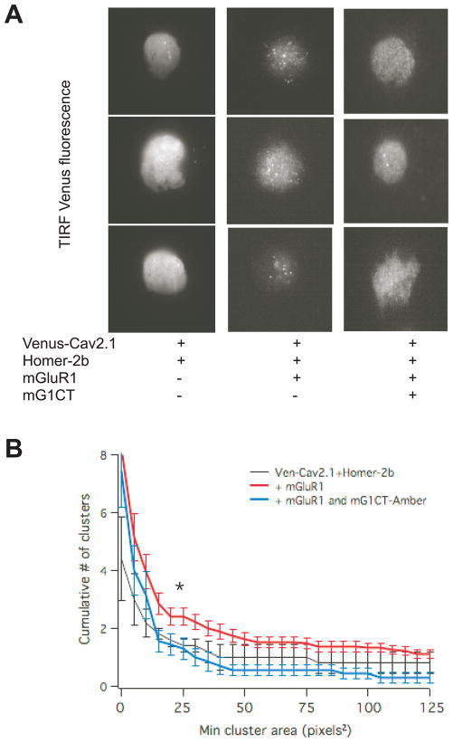
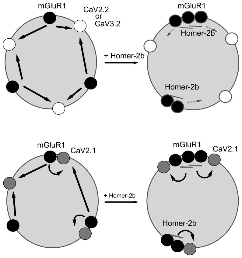
Group I metabotropic glutamate receptors (mGluR1 and 5) are G protein coupled receptors that regulate neuronal activity in a number of ways. Some of the most well studied functions of group I mGluRs, such as initiation of multiple forms of mGluR-dependent long-term depression, require receptor localization near the post-synaptic density (PSD). This localization is in turn dependent on the Homer family of scaffolding proteins which bind to a small motif on the distal C-termini of mGluR1 and 5, localize the receptors near the PSD, strengthen coupling to post-synaptic effectors and simultaneously uncouple the mGluRs from extra-synaptic effectors such as voltage dependent ion channels. Here the selectivity of this uncoupling process was examined by testing the ability of Homer-2b to uncouple mGluR1 from multiple voltage dependent calcium channels including Ca(V2.2) (N-type), Ca(V3.2) (T-type), and Ca(V2.1) (P/Q-type) expressed in rat sympathetic neurons from the superior cervical ganglion (SCG). Of these, only the mGluR1-Ca(V2.1) modulatory pathway was insensitive to Homer-2b expression. Uncoupling from this channel was achieved by co-expression of an mGluR1 C-terminal protein designed to disrupt a previously described direct interaction between these two proteins, suggesting that this interaction allows incorporation of Ca(V2.1) into the mGluR1/Homer signaling complex, thereby preserving modulation in the presence of scaffolding Homer proteins. This article is part of a Special Issue entitled 'Metabotropic Glutamate Receptors'.


Figures

Fig. 1
Fig. 1
CaV2.1 co-immunoprecipitates with mGluR1 in HEK293 cells. A, Anti-CaV2.1 (1:500) immunoblot showing protein from supernatant (S), flow-through (FT) and from the anti-HA immunoprecipitation (1:500) experiment (IP), with expression conditions indicated above: CaV2.1 alone and HA-mGluR1 + CaV2.1 (left) and HA-mGluR1 alone (right). B, Similar to A, but using an anti-myc antibody paired with myc-mGluR1 for the IP. Arrow indicates 250 kd marker.
Fig. 2
Fig. 2
CaV2.1 expression in SCG neurons. A, Sample current density time courses from cells expressing mGluR1 alone (filled circles), or mGluR1 plus CaV2.1 (open circles). Current inhibition by 100 μM glutamate (Glu) is evident under both conditions. CTX/SNX indicates application of 1 μM ω-conotoxin GVIA and 500 nM SNX482. Current measurements were obtained 10 msec into a test pulse to +10 mV. B, Sample current traces from the same cells as in A. The voltage protocol is shown above, and consisted of a 25 msec step from a −80 mV holding potential to + 10 mV. Control (uninhibited) currents are shown (con) as are currents maximally inhibited by the CTX-SNX toxin combination. C, Average (± SEM) current densities for SCG neurons expressing mGluR1 alone or mGluR1 plus CaV2.1. Asterisk indicates that current densities are significantly different (T-test, p < 0.05).
Fig. 3
Fig. 3
CaV2.1 current modulation by mGluR1 is insensitive to Homer protein expression. A, current amplitude time courses showing modulation by glutamate (Glu) of the total current and the isolated CaV2.1 current (in the presence of CTS/SNX) for SCG neurons expressing mGluR1 and CaV2.1 (upper) or mGluR1, CaV2.1, and Homer-2b (lower). B, average (± SEM) calcium current inhibition by 100 μM glutamate of the total current (left) and isolated CaV2.1 current (right) for neurons with the indicated expression. Number of cells for each condition shown in parentheses.
Fig. 4
Fig. 4
Overexpression of other voltage dependent calcium channels in SCG neurons does not preserve coupling to mGluR1 in the presence of Homer-2b. A, sample current traces (left) and time courses (right) illustrating the two-step voltage protocol (top) used to separate low voltage activated (CaV3.2) currents (LVA) from the native high voltage activated currents (HVA), and inhibition by 100 μM glutamate. Uninjected SCG neurons (upper left) showed no LVA currents. Neurons injected with mGluR1 and CaV3.2 (middle left and upper right) had prominent LVA and HVA currents, both of which were modulated by 100 μM glutamate (Glu). Additional co-expression of Homer-2b (lower left and lower right) uncoupled both the LVA and HVA currents from glutamate modulation. B, Average (± SEM) LVA and HVA calcium current inhibition by 100 μM glutamate in cells with the indicated expression. Number of cells is shown in parentheses. C, Average (± SEM) calcium current inhibition by 100 μM glutamate in control cells (expressing mGluR1, CaV2.2, and accessory proteins) and in cells co-expressing Homer-2b. Asterisk indicates a statistically significant difference from paired control (without Homer-2b; p ≤ 0.05). Number of cells is shown in parentheses.
Fig. 5
Fig. 5
CaV2.1 current modulation by mGluR1 is rendered sensitive to Homer protein expression when an mGluR1-CT construct is co-expressed to prevent the receptor-channel interaction. A, Current amplitude time courses showing modulation by glutamate (Glu) of the total current and the isolated CaV2.1 current (in the presence of CTS/SNX) for SCG neurons expressing mGluR1, mG1-CT and CaV2.1 (upper) or mGluR1,, mG1-CT, CaV2.1, and Homer-2b (lower). B, average (± SEM) calcium current inhibition by 100 μM glutamate of the total current (left) and isolated CaV2.1 current (right) for neurons with the indicated expression. Number of cells for each condition shown in parentheses. Asterisk indicates a statistically significant difference from paired control (without Homer-2b; p ≤ 0.05).
Fig. 6
Fig. 6
Coexpression of mGluR1 and Homer-2b induces Venus-CaV2.1 clustering, which is reduced by expression of an untagged mGluR1-CT construct. A, TIRF fluorescence images showing three sample images each of SCG neurons with the indicated expression. B, Clusters were quantified with a cumulative histogram of cluster number vs. size (in pixels2). * indicates statistically significant difference between the number of clusters in cells expressing Venus-CaV2.1, mGluR1 and Homer-2b (red; n=19) compared to cells also expressing mGluR1-CT-Amber (blue; n=7). Cluster histogram for neurons expressing Ven-CaV2.1 + Homer-2b (n=5) are shown in black. Lines represent average clusters ± SEM at 5 pixels2 bins.
Fig. 7
Fig. 7
Revised model describing the regulation of mGluR signaling by Homer proteins. Upper model shows the traditional model in which unclustered, Homer-free mGluR1 couples strongly to voltage dependent calcium channels (CaV2.2 and CaV3.2). Interaction with scaffolding Homer proteins (Homer-2b) results in mGluR1 clustering and uncoupling from channel modulation. Lower model shows revised scenario in which interaction with and coupling to CaV2.1 is preserved in the presence and absence of clustering and Homer expression.

References

    1. Ango F, Pin JP, Tu JC, Xiao B, Worley PF, Bockaert J, Fagni L. Dendritic and axonal targeting of type 5 metabotropic glutamate receptor is regulated by homer1 proteins and neuronal excitation. Journal of Neuroscience. 2000;20:8710–8716. - PMC - PubMed
    1. Brakeman PR, Lanahan AA, O’Brien R, Roche K, Barnes CA, Huganir RL. Worley P.F. Homer: a protein that selectively binds metabotropic glutamate receptors. Nature. 1997;386:284–288. - PubMed
    1. Campbell KP, Leung AT, Sharp AH. The biochemistry and molecular biology of the dihydropyridine-sensitive calcium channel. Trends in Neurosciences. 1988;11:425–430. - PubMed
    1. Catterall WA. Voltage-gated calcium channels. Cold Spring Harb Perspect Biol. 2011;3:a003947. - PMC - PubMed
    1. Chemin J, Traboulsie A, Lory P. Molecular pathways underlying the modulation of T-type calcium channels by neurotransmitters and hormones. Cell Calcium. 2006;40:121–134. - PubMed

MeSH terms

Substances