. 2012 Aug 15;31(16):3411-21.
doi: 10.1038/emboj.2012.157. Epub 2012 Jun 1.

Alternating access mechanism in the POT family of oligopeptide transporters

Affiliations

Alternating access mechanism in the POT family of oligopeptide transporters

Nicolae Solcan et al. EMBO J. .

Abstract

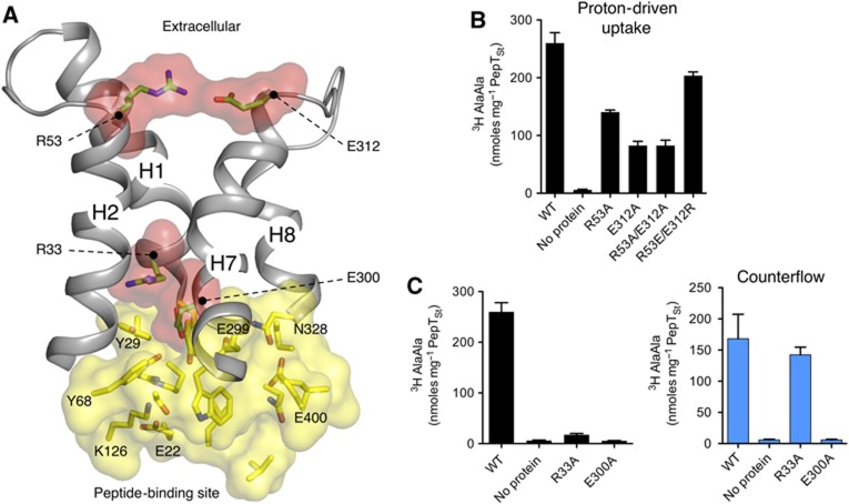
Short chain peptides are actively transported across membranes as an efficient route for dietary protein absorption and for maintaining cellular homeostasis. In mammals, peptide transport occurs via PepT1 and PepT2, which belong to the proton-dependent oligopeptide transporter, or POT family. The recent crystal structure of a bacterial POT transporter confirmed that they belong to the major facilitator superfamily of secondary active transporters. Despite the functional characterization of POT family members in bacteria, fungi and mammals, a detailed model for peptide recognition and transport remains unavailable. In this study, we report the 3.3-Å resolution crystal structure and functional characterization of a POT family transporter from the bacterium Streptococcus thermophilus. Crystallized in an inward open conformation the structure identifies a hinge-like movement within the C-terminal half of the transporter that facilitates opening of an intracellular gate controlling access to a central peptide-binding site. Our associated functional data support a model for peptide transport that highlights the importance of salt bridge interactions in orchestrating alternating access within the POT family.


Conflict of interest statement

The authors declare that they have no conflict of interest.

Figures

Figure 1
Figure 1
PepTSt structure reveals an inward open conformation. (A) Overall structure of PepTSt viewed from the extracellular side of the molecule. The 12-TM MFS fold is coloured blue to red with helices HA and HB coloured grey. The helices are labelled. The right-hand image shows a view in the plane of membrane with approximate dimensions of the molecule. (B) Slab through the surface electrostatic potential of PepTSt viewed in the plane of membrane to highlight the extracellular gate, central peptide-binding site and intracellular gate. (C) Peptide transport by PepTSt is driven by an inwardly directed proton gradient and selective for L di- and tri-peptides. ‘No competitor’ refers to the condition where only 3H di-alanine peptide was present in the external buffer, without additional cold peptides. ‘No protein’ refers to empty liposomes. ‘CCCP’ refers to addition of the proton ionophore carbonyl cyanide m-chlorophenyl hydrazine to the external buffer. Error bars indicate the standard deviations from triplicate experiments. (D) PepTSt displays higher affinity for hydrophobic di-peptides and can discriminate based on peptide size and charge.
Figure 2
Figure 2
Salt bridges facilitate closure of the extracellular gate. (A) Two prominent salt bridge interactions (Arg53–Glu312: distance ∼2.9 Å and Arg33–Glu300: distance ∼3.8 Å) are observed facilitating the close packing between helix hairpins H1–H2 and H7–H8 in the extracellular gate. Residues forming the central peptide-binding site are shown in yellow and the salt bride interactions in red. Helices and residues are labelled. (B) Effect of mutations in the Arg53–Glu312 salt bridge on proton-driven 3H di-alanine uptake. (C) Effect of mutations in the Arg33 and Glu300 salt bridge on proton driven (left-black bars) and peptide-driven counterflow transport (right-blue bars). Details of the counterflow experiments can be found in Supplementary data. Error bars indicate the standard deviations from triplicate experiments.
Figure 3
Figure 3
Proton binding and peptide specificity reside predominantly within the N-terminal domain of PepTSt. (A) Peptide-binding site as viewed from the plane of the membrane showing the ExxERFxYY motif on helix H1. Residues are coloured according to their predicted role, with proton binding (green), peptide specificity (pink) and transport (yellow). (B) Effect of substitutions within the peptide-binding site on proton-driven uptake. (C) Effect of equivalent substitutions on peptide driven counterflow uptake. (D) Effect of phenylalanine and alanine substitutions at Tyr29 and Tyr68 on the ability of different L-isomer peptides to compete for uptake of di-alanine in proton-driven uptake. (E) Change in IC50 values upon substitution of Tyr29 and Tyr68 to phenylalanine. The IC50 values for the different peptides were calculated as described in Supplementary data. Error bars indicate the standard deviations from triplicate experiments.
Figure 4
Figure 4
The intracellular gate is controlled through localized hinge bending in helices H10 and H11. (A) Comparison between the inward open PepTSt (magenta) and occluded PepTSo structure (grey) (PDB 2XUT) with arrows showing the hinge-like movement that opens the intracellular gate. View is from the membrane plane. The intracellular gate is shown in the occluded (PepTSo) and open (PepTSt) states. Residue numbers are for PepTSt. The peptide-binding site containing Lys126 and Glu400 is indicated. Right, view rotated 90° showing the Gly407 and Trp427 as spheres and their position within the helices. (B) Effect of mutating the hinge residues on proton driven (left-black bars) and peptide-driven counterflow uptake (right-blue bars) in PepTSt. Error bars indicate the standard deviations from triplicate experiments.
Figure 5
Figure 5
A model for proton-driven peptide symport by PepTSt. (A) PepTSt adopts an outward facing state, here modelled on the outward facing fucose permease structure (PDB: 3O7Q). This state is characterized by the packing of helices H4, H5 with H10, H11 that form the intracellular gate and is potentially stabilized through a salt bridge interaction between K126 and E400 as discussed. Peptide (Pep) and proton (H+) bind from the extracellular side of the membrane with important roles for the N-terminal ExxERFxYY motif on H1 and K126 in H4. (B) Binding results in closure of the extracellular gate to form the occluded state, here modelled on the occluded structure of PepTSo (PDB: 2XUT). This conformation is characterized by the packing of helices H7, H8 against H1, H2 at the extracellular side of the binding site, assisted through the formation of the salt bridge interactions between R53-E312 and R33-E300. Binding of both peptide and proton is also likely to disrupt the proposed interaction between K126 and E400, thereby facilitating release of the intracellular gate. In the occluded state, an additional extracellular cavity may also form in the C-terminal domain following closure of the extracellular gate, as observed in the occluded PepTSo structure. (C) Transition to the inward facing state, modelled on the inward facing PepTSt structure (PDB: 4APS) occurs in part through a localized hinge-like movement in helices H10, H11 that results in release of the intracellular gate, allowing exit of proton and peptide into the interior of the cell.

Comment in

  • A POTluck of peptide transporters.
    Nissen P. Nissen P. EMBO J. 2012 Aug 15;31(16):3382-3. doi: 10.1038/emboj.2012.206. Epub 2012 Jul 27. EMBO J. 2012. PMID: 22842786 Free PMC article.

References

    1. Abrahams JP, Leslie AGW (1996) Methods used in the structure determination of bovine mitochondrial F1 ATPase. Acta Crystallogr D Biol Crystallogr 52: 30–42 - PubMed
    1. Abramson J, Smirnova I, Kasho V, Verner G, Kaback HR, Iwata S (2003) Structure and mechanism of the lactose permease of Escherichia coli. Science 301: 610–615 - PubMed
    1. Anderson CMH, Thwaites DT (2010) Hijacking solute carriers for proton-coupled drug transport. Physiology 25: 364–377 - PubMed
    1. Biegel A, Knutter I, Hartrodt B, Gebauer S, Theis S, Luckner P, Kottra G, Rastetter M, Zebisch K, Thondorf I, Daniel H, Neubert K, Brandsch M (2006) The renal type H+/peptide symporter PEPT2: structure-affinity relationships. Amino Acids 31: 137–156 - PubMed
    1. Blanc E, Roversi P, Vonrhein C, Flensburg C, Lea SM, Bricogne G (2004) Refinement of severely incomplete structures with maximum likelihood in BUSTER-TNT. Acta Crystallogr D Biol Crystallogr 60: 2210–2221 - PubMed

Publication types

MeSH terms

Substances

Associated data