Review
. 2012 Mar-Apr;6(2):148-56.
doi: 10.4161/cam.20888. Epub 2012 Mar 1.

Bone cell interactions through Eph/ephrin: bone modeling, remodeling and associated diseases

Affiliations
Review

Bone cell interactions through Eph/ephrin: bone modeling, remodeling and associated diseases

Koichi Matsuo et al. Cell Adh Migr. 2012 Mar-Apr.

Abstract

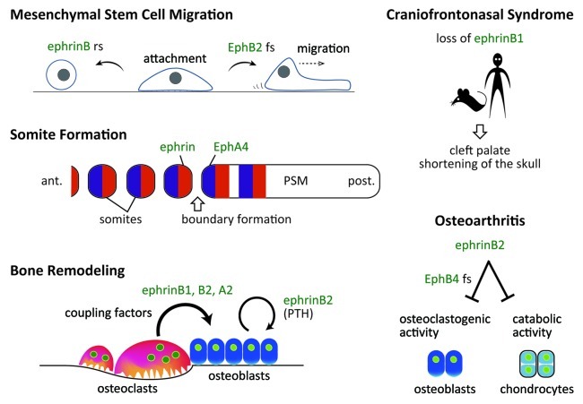
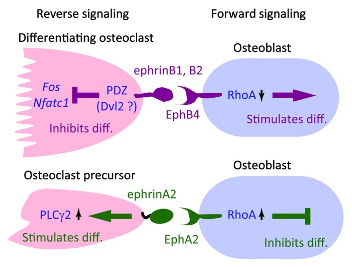
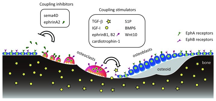
Bones cannot properly form or be maintained without cell-cell interactions through ephrin ligands and Eph receptors. Cell culture analysis and evaluation of genetic mouse models and human diseases reveal various ephrins and Eph functions in the skeletal system. Migration, attachment and spreading of mesenchymal stem cells are regulated by ephrinB ligands and EphB receptors. ephrinB1 loss-of-function is associated with craniofrontonasal syndrome (CFNS) in humans and mice. In bone remodeling, ephrinB2 is postulated to act as a "coupling stimulator." In that case, bidirectional signaling between osteoclastic ephrinB2 and osteoblastic EphB4 suppresses osteoclastic bone resorption and enhances osteoblastic bone formation, facilitating the transition between these two states. Parathyroid hormone (PTH) induces ephrinB2 in osteoblasts and enhances osteoblastic bone formation. In contrast to ephrinB2, ephrinA2 acts as a "coupling inhibitor," since ephrinA2 reverse signaling into osteoclasts enhances osteoclastogenesis and EphA2 forward signaling into osteoblasts suppresses osteoblastic bone formation and mineralization. Furthermore, ephrins and Ephs likely modulate pathological conditions such as osteoarthritis, rheumatoid arthritis, multiple myeloma and osteosarcoma. This review focuses on ephrin/Eph-mediated cell-cell interactions in bone biology.


Figures

None
Figure 1. Ephrins and Ephs in bone biology (an overview). rs, reverse signaling; fs, forward signaling; ant., anterior; post., posterior; PSM, presomitic mesoderm.
None
Figure 2. Expression of ephrins and Ephs during bone cell differentiation. (A) Osteoclast differentiation is induced by RANKL and M-CSF in vitro and then ephrin/Eph mRNA expression is monitored on the days indicated. (B) Osteoblast differentiation is induced by ascorbic acid and β-glycerophosphate in vitro and ephrin/Eph mRNA expression was similarly analyzed. Bar height represents changes in relative expression levels of each gene, based mainly on RT-PCR data., Levels can be compared only within a gene, not across genes. ephrin and Eph names in bold are discussed in detail in this review. Asterisks (*) indicate that mRNA expression levels are accompanied by corresponding changes in protein expression.,,
None
Figure 3. Osteoclast-osteoblast interactions through ephrins/Ephs. ephrinB2 is expressed in differentiating and mature osteoclasts, while ephrinA2 is expressed in early differentiating osteoclasts., Reverse signaling through ephrinB2, which may be mediated by PDZ domain proteins such as Dishevelled 2 (Dvl2), suppresses transcription of Fos and Nfatc1, thereby inhibiting osteoclast differentiation. Reverse signaling through ephrinA2 is mediated by activation of phospholipase Cγ2 (PLCγ2). Forward signaling through EphB4 suppresses RhoA activity and thereby stimulates osteoblast differentiation, while signaling through EphA2 likely enhances RhoA activity and inhibits osteoblast differentiation., How RhoA activity is differentially regulated by EphB4 and EphA2 signaling is unclear.
None
Figure 4. Coupling stimulators and inhibitors during bone remodeling. Bone matrix contains TGF-β (yellow stars) and IGF-I (yellow circles), which are released by osteoclastic bone resorption to stimulate coupling. Cells in the osteoclast lineage (red) produce various coupling stimulators and inhibitors that act on osteoblasts or their progenitors (blue).

References

    1. Pasquale EB. Eph-ephrin bidirectional signaling in physiology and disease. Cell. 2008;133:38–52. doi: 10.1016/j.cell.2008.03.011. - DOI - PubMed
    1. Pitulescu ME, Adams RH. Eph/ephrin molecules--a hub for signaling and endocytosis. Genes Dev. 2010;24:2480–92. doi: 10.1101/gad.1973910. - DOI - PMC - PubMed
    1. Egea J, Klein R. Bidirectional Eph-ephrin signaling during axon guidance. Trends Cell Biol. 2007;17:230–8. doi: 10.1016/j.tcb.2007.03.004. - DOI - PubMed
    1. Davy A, Soriano P. Ephrin signaling in vivo: look both ways. Dev Dyn. 2005;232:1–10. doi: 10.1002/dvdy.20200. - DOI - PubMed
    1. Pasquale EB. Eph receptors and ephrins in cancer: bidirectional signalling and beyond. Nat Rev Cancer. 2010;10:165–80. doi: 10.1038/nrc2806. - DOI - PMC - PubMed

Publication types