. 2012 May 9;485(7400):661-5.
doi: 10.1038/nature11066.

AMPK regulates NADPH homeostasis to promote tumour cell survival during energy stress

Affiliations

AMPK regulates NADPH homeostasis to promote tumour cell survival during energy stress

Sang-Min Jeon et al. Nature. .

Abstract

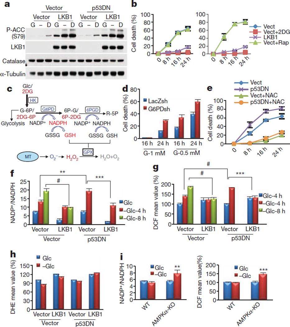
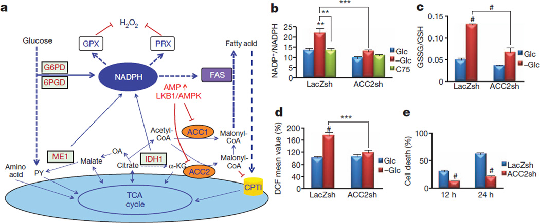
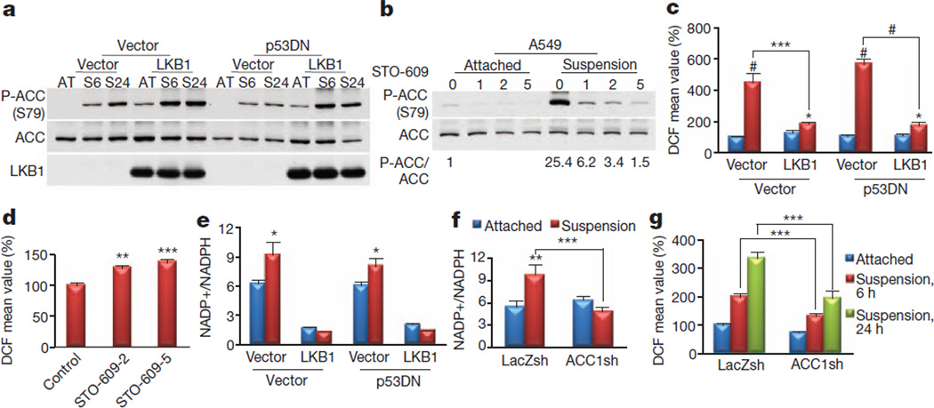
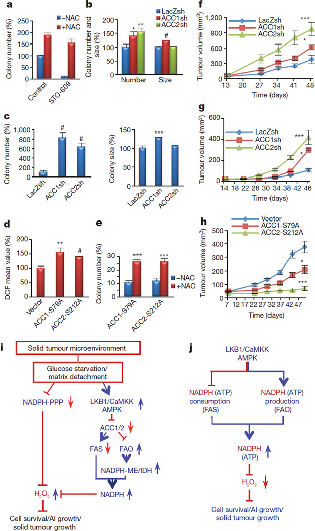
Overcoming metabolic stress is a critical step for solid tumour growth. However, the underlying mechanisms of cell death and survival under metabolic stress are not well understood. A key signalling pathway involved in metabolic adaptation is the liver kinase B1 (LKB1)-AMP-activated protein kinase (AMPK) pathway. Energy stress conditions that decrease intracellular ATP levels below a certain level promote AMPK activation by LKB1. Previous studies showed that LKB1-deficient or AMPK-deficient cells are resistant to oncogenic transformation and tumorigenesis, possibly because of the function of AMPK in metabolic adaptation. However, the mechanisms by which AMPK promotes metabolic adaptation in tumour cells are not fully understood. Here we show that AMPK activation, during energy stress, prolongs cell survival by redox regulation. Under these conditions, NADPH generation by the pentose phosphate pathway is impaired, but AMPK induces alternative routes to maintain NADPH and inhibit cell death. The inhibition of the acetyl-CoA carboxylases ACC1 and ACC2 by AMPK maintains NADPH levels by decreasing NADPH consumption in fatty-acid synthesis and increasing NADPH generation by means of fatty-acid oxidation. Knockdown of either ACC1 or ACC2 compensates for AMPK activation and facilitates anchorage-independent growth and solid tumour formation in vivo, whereas the activation of ACC1 or ACC2 attenuates these processes. Thus AMPK, in addition to its function in ATP homeostasis, has a key function in NADPH maintenance, which is critical for cancer cell survival under energy stress conditions, such as glucose limitations, anchorage-independent growth and solid tumour formation in vivo.


Figures

Figure 1
Figure 1. The failure to activate AMPK accelerates NADPH depletion, oxidative stress and cell death in the absence of glucose
a, Immunoblotting analyses after incubating A549 cells expressing empty vector, LKB1, p53DN or both p53DN and LKB1 in the absence (−) or presence of 5mMglucose (G) or 2DG (D) for 2 h. b, Cell death quantification after glucose starvation. For A549-Vect (left) and A549-p53DN (right) cells, 5 mM 2DG (Vect+2DG) or 100 nM rapamycin (Vect+Rap) were added to the medium. c, Illustration depicting partial metabolism of 2DG through the PPP, which could generate NADPH/GSH and eliminate H2O2 generated from mitochondria (MT) (see the text for details). Glc, glucose; HK, hexokinase; G-6P, glucose 6-phosphate; 6PGD, 6-phosphogluconate dehydrogenase; 2DG-6P, 2-deoxyglucose 6-phosphate; 6P-G, 6-phosphogluconate; 6P-2DG, 6-phospho 2-deoxygluconate; R-5P, ribulose 5-phosphate. d, Quantification of cell death of A549 cells expressing LacZ-shRNA or G6PD-shRNA cultured in 1 mM or 0.5 mM glucose. e, Cell death quantification at different time points after glucose depletion in the presence or absence of 2 mM NAC. f, g, NADP+/NADPH ratio (f) and H2O2 level (g) after incubation in the absence (−Glc) or presence of 5 mM glucose (Glc) for 4 or 8 h. h, O2 levels after incubation in the absence or presence of 5 mM glucose for 4 h. i, NADP+/NADPH ratio (left) and H2O2 level (right) in wild-type (WT) or AMPKα-KO MEFs cultured in glucose-free medium in the absence (−Glc) or presence of 5 mM glucose (Glc) for 5 h. Results in g, h and i (right) are expressed as the percentage change in the mean DCF/DHE values relative to the glucose-treated control. Results are shown as means and s.e.m. for three independent (b, d, f, h, i) or four (e, g) experiments. Asterisk, P < 0.05; two asterisks, P < 0.01; three asterisks, P < 0.005; hash, P < 0.001 versus the glucose-treated control in each group.
Figure 2
Figure 2. ACC2 ablation recapitulates AMPK activation, maintaining the NADPH level and decreasing the H2O2 level during glucose deprivation
a, A schematic illustration depicting one potential mechanism by which AMPK regulates NADPH homeostasis (see the text for details). PY, pyruvate; OA, oxaloacetate; α-KG, α-oxoglutarate; ME, malic enzyme; IDH, isocitrate dehydrogenase; TCA, tricarboxylic acid. b–d, A549 cells expressing LacZ-shRNA or ACC2-shRNA 2 were incubated in glucose-free medium in the absence (−Glc) or presence of 5 mM glucose (Glc) or with 20 µg ml−1 C75. After 6 h, the NADP+/NADPH ratio (b), GSSG/GSH ratio (c) and H2O2 level (d) were measured. e, Quantification of cell death after glucose starvation for 10 and 24 h. Results in d are expressed as the percentage change in the mean DCF values relative to the glucose-treated control. Results are shown as means and s.e.m. for three independent experiments. Two asterisks, P < 0.01; three asterisks, P < 0.005; hash, P < 0.001 versus the glucose-treated control in each group (b–d) or versus the LacZsh control (e) at each time point.
Figure 3
Figure 3. AMPK-mediated inhibition of ACC1 is required to maintain the NADPH and reactive oxygen species levels after matrix detachment
a, A549-Vect-Vect, A549-Vect-LKB1, A549-p53DN-Vect and A549-p53DN-LKB1 cells were grown attached (AT) or in suspension (S6, S24) and subjected to immunoblotting at the indicated time points (6 and 24 h). b, A549 cells were grown attached or in suspension in the presence or absence of STO-609 (1, 2 or 5 µg ml−1) for 6 h and subjected to immunoblotting for quantification of phospho-ACC (P-ACC). c, d, H2O2 levels after growth attached (blue columns) or in suspension (red columns) for 24 h (c) or for 6 h in the presence or absence of 2 or 5 µg ml−1 STO-609 (d). e, NADP+/NADPH ratio after growth attached (blue columns) or in suspension (red columns) for 24 h. f, g, The effect of ACC knockdown on NADP+/NADPH ratio (f) or H2O2 level (g) after in suspension for 24 h (f) or for 6 and 24 h (g). Results in c, d and g are expressed as the percentage change in the mean DCF values over the values of the attached control. Results are shown as means and s.e.m. for three independent experiments. Asterisk, P < 0.05; two asterisks, P < 0.01; three asterisks, P < 0.005; hash, P < 0.001 versus the attached condition in each group (c, e–g) or versus the control (d).
Figure 4
Figure 4. AMPK-mediated inhibition of ACC1 or ACC2 promotes anchorage-independent growth and solid tumour formation in vivo
a, Quantification of soft agar colonies of A549 cells in the presence or absence of 5 µg ml−1 STO-609, with or without 2 mM NAC. b, c, A549 cells (b) or AMPKα-KO-RasV12-MEFs (c) expressing LacZ-shRNA, ACC1-shRNA, or ACC2-shRNA were plated on soft agar, and the number and size of the colonies were analysed. Results are expressed as the percentage change in the colony number and size relative to the −NAC control (a) or the LacZsh control (b, c). d, e, MCF7 cells expressing ACC1-S79A or ACC2-S212A were generated and grown in suspension for 6 h before quantification of the H2O2 level (d) or were subjected to soft agar assay (with or without 2 mM NAC) (e). Results are expressed as the percentage change in the mean DCF values (d) or colony number (e) over the values of the vector control. Results are expressed as means and s.e.m. for three independent experiments. f, g, A549 cells (f), or AMPKα-KO-RasV12 cells (g) expressing LacZ-shRNA, ACC1-shRNA, or ACC2-shRNA were injected subcutaneously into nude mice. h, MCF7 cells expressing vector, ACC1-S79A or ACC2-S212A were mixed with Matrigel (10%) and injected orthotopically into the mammary fat pads of nude mice. Tumour growth was monitored and measured weekly. Results are shown as means and s.e.m. for four mice in each group. Asterisk, P < 0.05; two asterisks, P < 0.01; three asterisks, P < 0.005; hash, P < 0.001 versus LacZsh (b, c), vector (d) or −NAC in each group (e), and versus LacZ-shRNA (f, g) or vector (h) at each time point. i, Summary: under energy stress when PPP is impaired, AMPK activation attenuates cell death by maintaining NADPH level through FAO-induced NADPH production and by inhibiting NADPH consumption in FAS. AI growth, anchorage-independent growth. j, Proposed model: in addition to its function in ATP homeostasis, the LKB1/CaMKK–AMPK/ACC1/2 axis has a key function in NADPH homeostasis to decrease H2O2 level and to promote cancer cell survival and metabolic adaptation.

Comment in

References

    1. Folkman J. Angiogenesis and apoptosis. Semin. Cancer Biol. 2003;13:159–167. - PubMed
    1. Jones RG, Thompson CB. Tumor suppressors and cell metabolism: a recipe for cancer growth. Genes Dev. 2009;23:537–548. - PMC - PubMed
    1. Shackelford DB, Shaw RJ. The LKB1–AMPK pathway: metabolism and growth control in tumour suppression. Nature Rev. Cancer. 2009;9:563–575. - PMC - PubMed
    1. Bardeesy N, et al. Loss of the Lkb1 tumour suppressor provokes intestinal polyposis but resistance to transformation. Nature. 2002;419:162–167. - PubMed
    1. Kato K, et al. Critical roles of AMP-activated protein kinase in constitutive tolerance of cancer cells to nutrient deprivation and tumor formation. Oncogene. 2002;21:6082–6090. - PubMed

Publication types

MeSH terms