. 2012 Jul 1;189(1):381-92.
doi: 10.4049/jimmunol.1200409. Epub 2012 Jun 1.

Neutrophil cerebrovascular transmigration triggers rapid neurotoxicity through release of proteases associated with decondensed DNA

Affiliations

Neutrophil cerebrovascular transmigration triggers rapid neurotoxicity through release of proteases associated with decondensed DNA

Charlotte Allen et al. J Immunol. .

Abstract

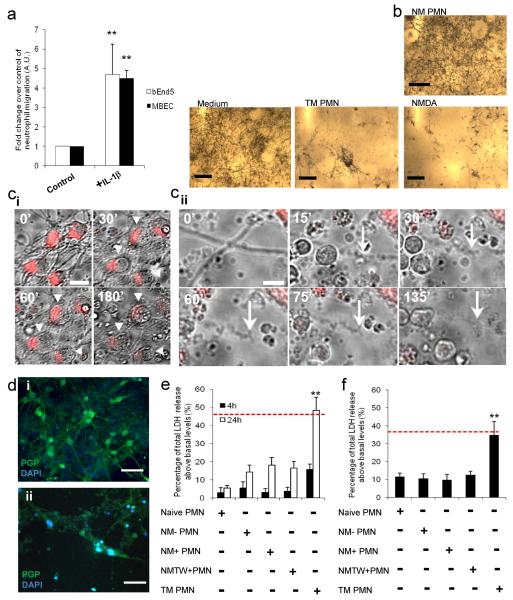
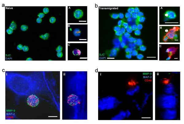
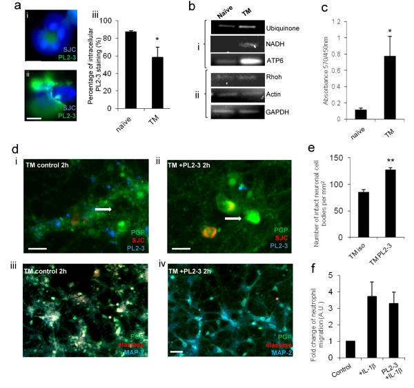
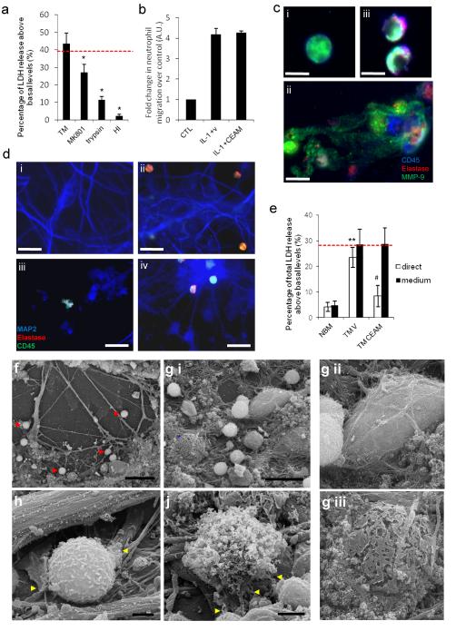
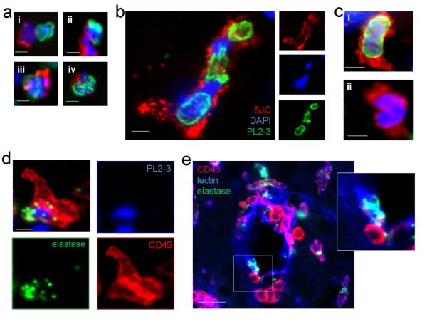
Cerebrovascular inflammation contributes to diverse CNS disorders through mechanisms that are incompletely understood. The recruitment of neutrophils to the brain can contribute to neurotoxicity, particularly during acute brain injuries, such as cerebral ischemia, trauma, and seizures. However, the regulatory and effector mechanisms that underlie neutrophil-mediated neurotoxicity are poorly understood. In this study, we show that mouse neutrophils are not inherently toxic to neurons but that transendothelial migration across IL-1-stimulated brain endothelium triggers neutrophils to acquire a neurotoxic phenotype that causes the rapid death of cultured neurons. Neurotoxicity was induced by the addition of transmigrated neutrophils or conditioned medium, taken from transmigrated neutrophils, to neurons and was partially mediated by excitotoxic mechanisms and soluble proteins. Transmigrated neutrophils also released decondensed DNA associated with proteases, which are known as neutrophil extracellular traps. The blockade of histone-DNA complexes attenuated transmigrated neutrophil-induced neuronal death, whereas the inhibition of key neutrophil proteases in the presence of transmigrated neutrophils rescued neuronal viability. We also show that neutrophil recruitment in the brain is IL-1 dependent, and release of proteases and decondensed DNA from recruited neutrophils in the brain occurs in several in vivo experimental models of neuroinflammation. These data reveal new regulatory and effector mechanisms of neutrophil-mediated neurotoxicity (i.e., the release of proteases and decondensed DNA triggered by phenotypic transformation during cerebrovascular transmigration). Such mechanisms have important implications for neuroinflammatory disorders, notably in the development of antileukocyte therapies.


Figures

Figure 1
Figure 1. Neutrophils acquire a neurotoxic phenotype after transmigration across activated brain endothelium
(a) Transmigration of neutrophils across brain endothelioma cells (bEnd.5) and primary murine brain endothelial cells (MBEC) treated with IL-1β (4 h) after 24 h. (b) Brightfield images of neurons exposed to neurobasal medium, non-migrated neutrophils (NM-PMN) or transmigrated neutrophils (TM PMN) (120,000 cells/cm2), or 600 μM NMDA in the presence of MTT (5 μg/ml) for 24 h. Neuronal viability under these conditions is indicated by the presence or absence of MTT crystals. (c) Brightfield and CellTracker Red images of stained neurons (arrowheads) exposed to TM PMN for 0-180 min (i, scale bar, 15 μm, and see movie 1). Brightfield images of a neuronal process (arrow) exposed to TM PMN for 0-135 min (ii, scale bar, 5 μm). (d) Immunofluorescent images of neurons (PGP, green) exposed to (i) naïve PMN or (ii) TM PMN (120,000 PMN /cm2) for 24 h (scale bar, 30 μm). (e) Quantification of total LDH release from neurons 4 h or 24 h after application of naïve neutrophils (PMN), non-migrated neutrophils, which had been incubated with non-activated endothelial cells (NM− PMN), non-migrated neutrophils, which had been incubated with IL-1β-activated brain endothelial cells (NM+ PMN), or incubated with conditioned medium from activated endothelium after washing, (NMTW+ PMN) or transmigrated neutrophils (TM PMN) (120,000 cells/cm2. (f) Effect of conditioned medium (applied at 1:4 dilution) from naive PMN, NM− PMN, NM+ PMN, NM+TW or TM PMN on neuronal viability as assessed by LDH release after 24 h. Bars represent the mean data +/− SEM for a minimum of 3 independent experiments carried out on separate cultures. Red dotted line indicates neuronal death induced by 600μM NMDA. **P<0.01 (one-way ANOVA, with a Bonferroni post hoc test).
Figure 2
Figure 2. Neutrophils migrated across IL-1β-activated cerebrovascular endothelium show altered phenotype in comparison to naïve controls
(a) Naïve neutrophils stained with (A) anti-neutrophil serum (SJC, green) and DAPI (blue), (B) SJC (red), DAPI (blue) and PL2-3 (green) or (C) elastase (red), DAPI (blue) and PL2-3 (green) (scale bar, 10 μm). (b) Transmigrated neutrophils stained with (A) SJC (green) and DAPI (blue), (B) SJC (red), DAPI (blue) and PL2-3 (green) or (C) elastase (red), DAPI (blue) and PL2-3 (green) (scale bar, 10 μm). Immunofluorescent images of neurons (MAP2, blue) exposed to (c i) naïve (z-stack c ii) or (d i) transmigrated neutrophils (z-stack d ii) (120,000 neutrophils /cm2) for 4 h. Neutrophils (CD45, red) contain high levels of MMP-9 (c i) (green), which is lost from transmigrated neutrophils added to neurons (d i) (scale bar, 5 μm).
Figure 3
Figure 3. Neutrophil proteases are associated with neutrophil extracellular traps and neuronal death in vitro
(a) Intracellular PL2-3 staining is significantly reduced in (ii) transmigrated (TM) neutrophils compared to (i) naïve neutrophils (DAPI and PL2-3) (scale bar, 5 μm), (iii) graph showing the percentage of neutrophils present with intracellular PL2-3 staining. (b) Neutrophil-derived mitochondrial DNA is increased in conditioned medium from transmigrated neutrophils but not in that of naïve neutrophils as determined by PCR. (i) mitochondrial and (ii) nuclear DNA was observed in the conditioned medium of transmigrated neutrophils compared to naïve controls. (c) Elevated levels of extracelluar histone-DNA was detected in conditioned medium of naïve neutrophils and transmigrated neutrophils, (d) Neurons were incubated for 2 h and stained with PGP (green), neutrophils were stained with SJC (red) and NETs stained with PL2-3 (blue) (scale bar, 10 μm). (iii) Reduction in neurotoxicity of transmigrated neutrophils in the presence of PL2-3 (15 μg/ml) is seen in neuronal cultures stained with PGP (green) and MAP-2 (red), as compared to the (iv) isotype treated control (scale bars, 30 μm). Maintained structural integrity is shown in cultured neurons (white arrows) after (ii) exposure to transmigrated neutrophils treated with PL2-3 (15 μg/ml) in comparison to (i) neutrophils transmigrated across activated endothelium in the presence of an isotype control (scale bar, 10 μm). (e) Percentage of maintained integrity of neuronal cell bodies after exposure to transmigrated neutrophils treated with PL2-3 (15 μg/ml) in comparison to isotype treated control.(f) Presence of PL2-3 antibody (15 μg/ml) does not affect neutrophil migration across IL-1β-activated brain endothelium after 24 h of migration. Bars in graphs represent the mean +/− SEM from a minimum of 3 independent experiments carried out on separate cultures. *P<0.05 **P<0.01 (Student’s t-test or one-way ANOVA, with a Bonferroni post hoc test or Student’s t-test where appropriate).
Figure 4
Figure 4. Degranulation and NET release are associated with neurotoxicity in transmigrated neutrophils through neutrophil proteases
(a) Neuronal LDH release is shown 24 h after treatment with conditioned medium from transmigrated (TM) neutrophils with or without pre-treatment of neurons with MK-801 (10 μM), trypsin or heat-inactivated (HI) CM. (b) The transmigration of neutrophils across IL-1β-activated brain endothelium (bEnd.5) is not significantly altered by the presence of CEAM after 24 h. (c) Neutrophil morphology indicated by presence of elastase (red), CD45 (blue) and MMP-9 (green) using immunofluorescence in (i) naïve neutrophils. (iii) shows an attenuation of degranulation in the presence of CEAM, when compared to the vehicle treated transmigrated neutrophils (ii) (scale bar, 5 μm). (d) Maintained neuronal structural integrity shown in neuronal cultures after exposure to transmigrated neutrophils in the presence of CEAM (iv) in comparison to (iii) neutrophils transmigrated across activated endothelium. Vehicle treated transmigrated neutrophils induced extensive neuronal damage when comparing with naive neutrophils. Neurons were incubated for 4 h and stained with MAP2 indicating neuronal viability (blue); neutrophils were stained with CD45 (green) and elastase (red) (scale bar, 30 μm). (e) Neuronal viability was established using LDH assay after the direct application of naïve and transmigrated neutrophils (white bars) or naïve and transmigrated neutrophils conditioned medium (black bars) in the presence of CEAM or a vehicle control (f) Scanning electron micrograph images of transmigrated neutrophils in primary murine neuronal cultures after 1 h incubation. The presence of neutrophils indicated by red arrows (scale bar 30 μm). (g) (i) Identification and comparison of a healthy neuronal body (white asterisk) with a dying neuronal body (blue asterisk) (scale bar 20 μm). Neuronal bodies shown in a higher magnification on (ii) and (iii). (h) Transmigrated neutrophil in initial phases of degranulation (yellow arrows) surrounding neuronal processes (scale bar 2 μm). (j) Full degranulation of the transmigrated neutrophils, where the granules and globular structures of NETs are clearly visualized (scale bar 2 μm). Bars represent the mean data +/− SEM for a minimum of 3 independent experiments carried out on separate cultures. Red dotted line indicates neuronal death induced by 600μM NMDA. *P<0.05 vs. TM; **P<0.01 TM vs. naïve (NBM); #P<0.05 TM vs. TM CEAM (one-way ANOVA, with Bonferroni's post-hoc test).
Figure 5
Figure 5. Neutrophil recruitment in acute brain injury is associated with extracellular proteases and loss of intracellular de-condensed DNA in vivo
(a) Loss of intracellular histone-DNA complexes (PL2-3 staining) in neutrophils identified in the cerebral cortex in vivo in different established models of neuroinflammation (i) after induction of cerebral ischaemia, (ii) after induction of cerebral ischaemia in the presence of peripherally injected IL-1β, (iii) after induction of cerebral ischaemia with following systemic infection with Trichuris muris (3), and (iv) after stereotaxic intrastriatal administration of IL-1 and AMPA in rat (scale bars, 5 μm). (b) In a population of recruited neutrophils in the inflamed brain PL2-3 staining is uneven and de-condensed DNA is observed in close proximity to nuclear DNA (DAPI, blue) (c) (i) Meningeal neutrophils contain high amounts of intracellular histone-DNA complexes (PL2-3) after stereotaxic injection of LPS (scale bar 10 μm). (ii) Neutrophils recruited to the inflamed cerebral cortex contain less de-condensed DNA (PL2-3) after stereotaxic injection of LPS (scale bar 10 μm). (d) Extracellular PL2-3 and elastase are found in the cerebral cortex in vivo after stereotaxic injection of LPS (scale bar, 5 μm). (e) Immunofluorescence showing perivascular inflammatory cells (CD45, red) containing neutrophil elastase (green) adhering to the endothelial monolayer (lectin, blue) in the brain (scale bar, 20 μm).

References

    1. Allan SM, Tyrrell PJ, Rothwell NJ. Interleukin-1 and neuronal injury. Nat Rev Immunol. 2005;5:629–640. - PubMed
    1. Neumann J, Sauerzweig S, Ronicke R, Gunzer F, Dinkel K, Ullrich O, Gunzer M, Reymann KG. Microglia cells protect neurons by direct engulfment of invading neutrophil granulocytes: a new mechanism of CNS immune privilege. J Neurosci. 2008;28:5965–5975. - PMC - PubMed
    1. Denes A, Humphreys N, Lane TE, Grencis R, Rothwell N. Chronic systemic infection exacerbates ischemic brain damage via a CCL5 (regulated on activation, normal T-cell expressed and secreted)-mediated proinflammatory response in mice. J Neurosci. 2010;30:10086–10095. - PMC - PubMed
    1. McColl BW, Rothwell NJ, Allan SM. Systemic inflammation alters the kinetics of cerebrovascular tight junction disruption after experimental stroke in mice. J Neurosci. 2008;28:9451–9462. - PMC - PubMed
    1. Soehnlein O, Lindbom L. Phagocyte partnership during the onset and resolution of inflammation. Nat Rev Immunol. 2010;10:427–439. - PubMed

Publication types

MeSH terms