. 2012 Jun 1;26(11):1196-208.
doi: 10.1101/gad.189274.112.

SUMO-targeted ubiquitin E3 ligase RNF4 is required for the response of human cells to DNA damage

Affiliations

SUMO-targeted ubiquitin E3 ligase RNF4 is required for the response of human cells to DNA damage

Yili Yin et al. Genes Dev. .

Abstract

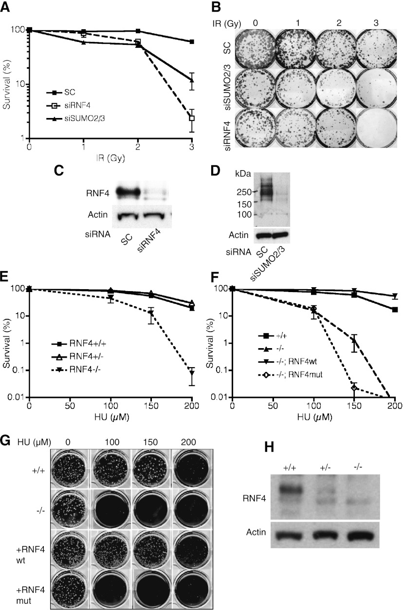
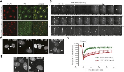
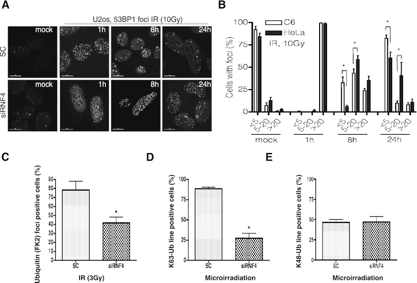
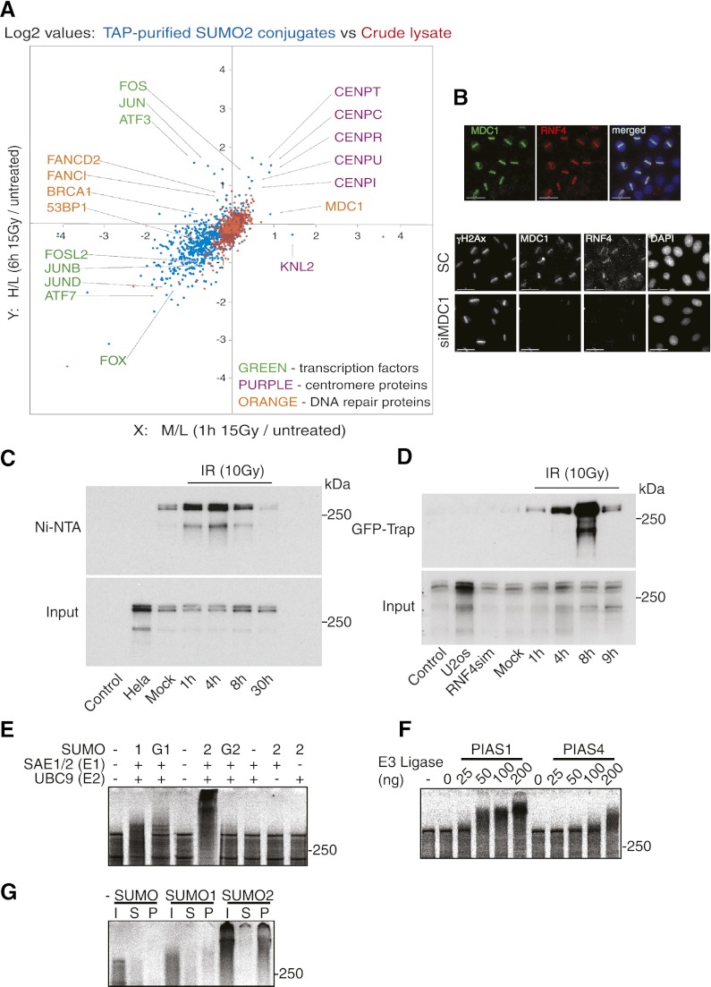
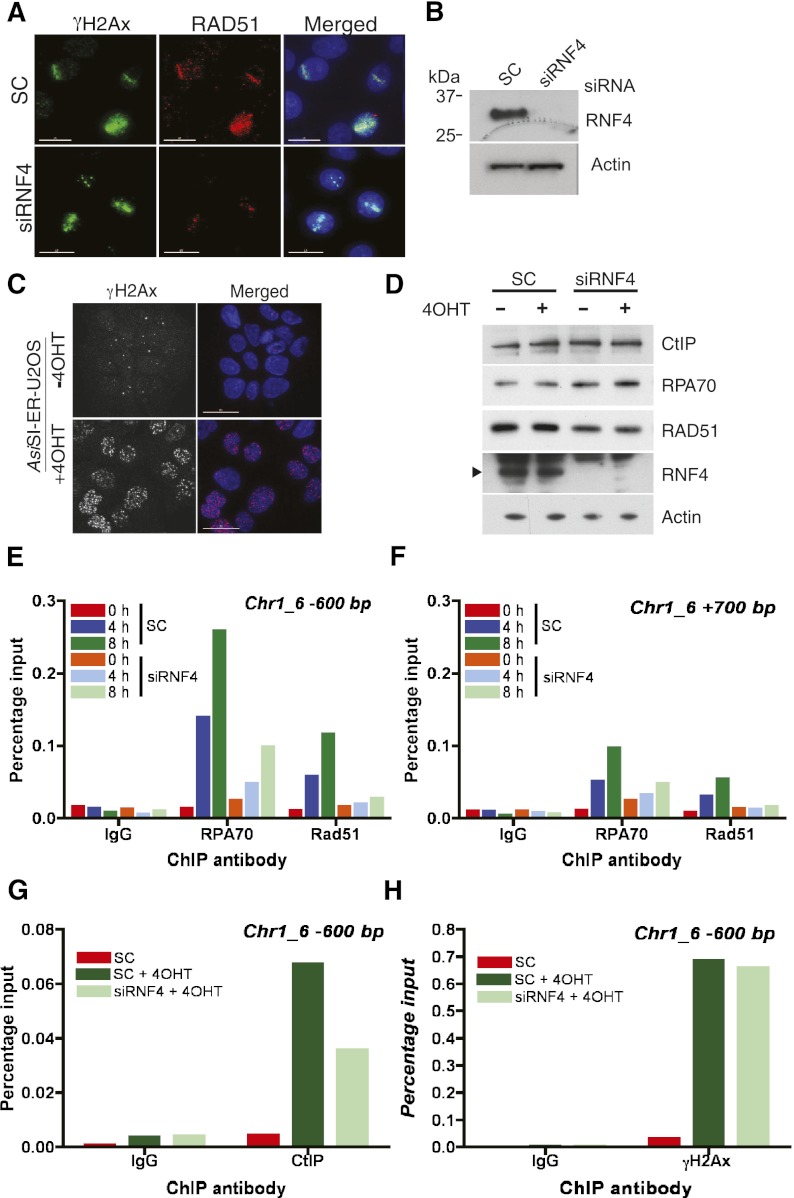
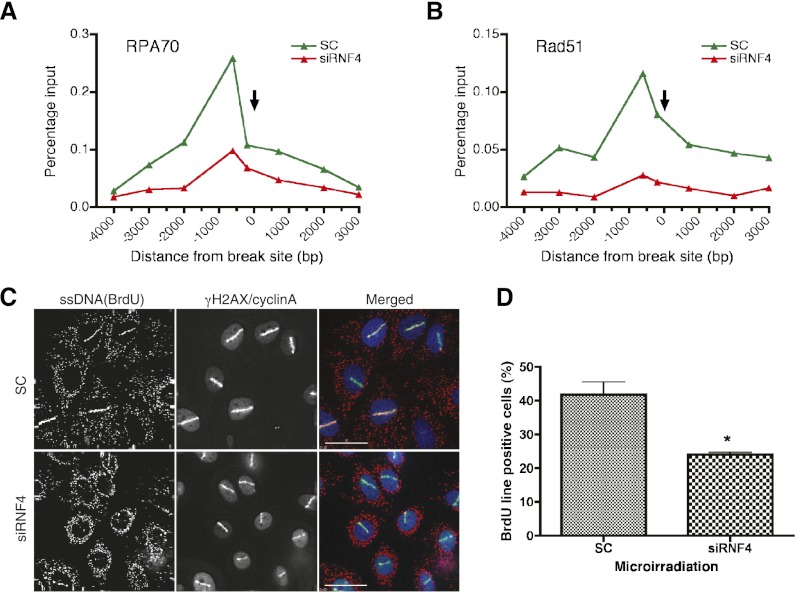
Here we demonstrate that RNF4, a highly conserved small ubiquitin-like modifier (SUMO)-targeted ubiquitin E3 ligase, plays a critical role in the response of mammalian cells to DNA damage. Human cells in which RNF4 expression was ablated by siRNA or chicken DT40 cells with a homozygous deletion of the RNF4 gene displayed increased sensitivity to DNA-damaging agents. Recruitment of RNF4 to double-strand breaks required its RING and SUMO interaction motif (SIM) domains and DNA damage factors such as NBS1, mediator of DNA damage checkpoint 1 (MDC1), RNF8, 53BP1, and BRCA1. In the absence of RNF4, these factors were still recruited to sites of DNA damage, but 53BP1, RNF8, and RNF168 displayed delayed clearance from such foci. SILAC-based proteomics of SUMO substrates revealed that MDC1 was SUMO-modified in response to ionizing radiation. As a consequence of SUMO modification, MDC1 recruited RNF4, which mediated ubiquitylation at the DNA damage site. Failure to recruit RNF4 resulted in defective loading of replication protein A (RPA) and Rad51 onto ssDNA. This appeared to be a consequence of reduced recruitment of the CtIP nuclease, resulting in inefficient end resection. Thus, RNF4 is a novel DNA damage-responsive protein that plays a role in homologous recombination and integrates SUMO modification and ubiquitin signaling in the cellular response to genotoxic stress.


Figures

Figure 1.
Figure 1.
Depletion of RNF4 sensitizes cells to genotoxic stress. (A,B) RNF4-depleted U2OS cells are sensitive to IR. U2OS cells were transfected with siRNA against SUMO-2/3 (siSUMO2/3), RNF4 (siRNF4), or a nontarget siRNA (SC) for 3 d and then exposed to IR at the indicated dose. Survival rates were determined by colony formation. The data represent the mean + SD (n = 3). (C,D) The efficiency of RNF4 or SUMO-2/3 depletion by siRNA in A and B was determined by Western blotting. (E–H) Chicken DT40 RNF4−/− cells display an increased sensitivity to genotoxic stress. Clonogenic survival assays were performed, and the number of surviving colonies was counted after chronic HU exposure. The assay was repeated with stably transfected clones expressing wild-type RNF4 (RNF4wt) or an E2-binding mutant of the rat ortholog of RNF4 (RNF4mut). The data represent the mean of three independent experiments, and the error bars indicate the SD. (H) Depletion of RNF4 in DT40 was confirmed by Western blotting (see also Supplemental Fig. S1).
Figure 2.
Figure 2.
RNF4 is recruited to sites of DNA damage. (A) Endogenous RNF4 colocalizes with γH2AX after DNA damage. HeLa cells or HeLa with RNF4 stably depleted (C6) were subjected to laser microirradiation. Immunofluorescence staining was performed using anti-RNF4 and anti-γH2AX antibodies. Specificity of the RNF4 antibody was confirmed by Western blotting after siRNA depletion. (B) Time-lapse experment showing the dynamic recruitment of RNF4. HeLa cells expressing YFP-RNF4 were laser-micro-irradiated, and redistribution of YFP-RNF4 was recorded for 13 h at 1-min intervals (Materials and Methods; see also Supplemental Movies S1, S2). (C) RNF4 recruitment to sites of micro-irradiation-induced DNA damage depends on its SIM and RING domains. HeLa cells were transfected with wild-type RNF4 (YFP-RNF4wt), SIM domain mutant RNF4 (YFP-RNF4sim), or a RING domain mutant (YFP-RNF4cs1) for 24 h and then laser-micro-irradiated. Time-lapse microscopy monitored recruitment of YFP-RNF4 for 2 h. The end point image is shown. Arrows indicate laser movement during micro-irradiation. (D) Quantitative FRAP analysis of YFP-RNFwt and YFP-RNF4cs1. The FRAP curves were derived from 20 cells for each condition. Normalized florescence intensity was compared. Error bars, SD. Data were analyzed by Student's t-test, and P-values were calculated (Materials and Methods; see also Supplemental Fig. S2). (E) RNF4 retention at sites of DNA damage requires SUMO modification. U2OS cells stably expressing YFP-RNF4 were transfected with SUMO-2/3-targeting siRNA or a nontarget siRNA control for 3 d, laser-micro-irradiated, and monitored for the recruitment of YFP-RNF4 along the laser track (arrows) using time-lapse microscopy. The image was derived from the 1-h time point. Bars: A, 30 μm; C,E, 15 μm.
Figure 3.
Figure 3.
RNF4 mediates repair and ubiquitin conjugation at sites of DNA damage. (A) RNF4 depletion delays DNA repair. U2OS cells were transfected with siRNA to RNF4 or a nontargeting siRNA (SC) for 3 d, exposed to 10 Gy of IR either prior to irradiation (mock) or 1, 8, or 24 h after irradiation, and immunostained with antibody against 53BP1 (see also Supplemental Fig. S4). (B) Quantification of data presented in Supplemental Figure S4c. At least 100 cells for each time point were scored. Cells were grouped by 53BP1 foci number: less than five, five to 20, and >20. Error bar, SD. (*) P < 0.05 (see also Supplemental Fig. S4c). (C) HeLa cells transfected with siRNA to RNF4 or a nontargeting siRNA (SC) for 72 h were exposed to 3 Gy of IR and allowed to recover for 1 h, and damage foci were visualized by staining with antibodies against γH2AX or conjugated ubiquitin (FK2). The percentage of γH2AX-positive cells that were also FK2-positive was determined; >100 cells were scored per condition. Data represent mean + SE from three independent experiments. (*) P < 0.05. (D) RNF4 mediates K63 ubiquitin chain formation after damage. Cells were treated with siRNA as in C and micro-irradiated before immunostaining using K63 ubiquitin-specific antibody and γH2AX antibody. The percentage of γH2AX-staining cells that were also stained for K63 ubiquitin along the laser track was calculated. More than 100 cells were scored per condition. Data represent mean + SE from two independent experiments. (*) P < 0.05. (E) Cells were treated with siRNA and micro-irridiation as in D. Immunostaining was performed using K48 ubiquitin-specific antibody. The percentage of cells with K48-Ub-positive laser tracks was determined as in D. More than 100 cells were scored per condition. Data represent mean + SE. (*) P < 0.05.
Figure 4.
Figure 4.
RNF4 binds to SUMO-modified MDC1 after DNA damage. (A) SILAC-based quantitative proteomic analysis (see the Materials and Methods) of SUMO modification after IR. TAP-SUMO-2 HeLa cells were grown in either normal medium, medium containing Lys4 and Arg6, or medium containing Lys8 and Arg10. Cells in normal medium were untreated, cells in Lys4 and Arg6 were exposed to 15 Gy of IR and harvested after 1 h, and cells in Lys8 and Arg10 were exposed to 15 Gy of IR and harvested after 6 h. Samples were mixed, and SUMO-modified proteins were analyzed by mass spectrometry. Crude lysates were also analyzed by mass spectrometry. (B) MDC1 is required for RNF4 recruitment to damage sites. The top panel shows that endogenous MDC1 and RNF4 colocalize after DNA damage. Laser-micro-irradiated HeLa cells were costained for endogenous MDC1 and RNF4. The bottom panel shows that RNF4 retention at DNA damage depends on MDC1. HeLa cells were treated with siRNA against MDC1 or a nontarget siRNA (SC) for 3 d, laser-micro-irradiated, and, after 1 h, immunostained with the indicated antibodies. Depletion of MDC1 by siRNA is shown in Supplemental Figures S6. (Bottom) Bars, 15 μm. (C) MDC1 is SUMO-modified after DNA damage in vivo. HeLa cells stably expressing His-SUMO2 were treated with or without 10 Gy of IR, collected at the indicated time point, and analyzed by Ni-NTA affinity chromatogaphy under denaturing condition, followed by Western blotting with MDC1-specific antibody. (D) RNF4 binds to MDC1 after DNA damage in vivo. U2OS stably expressing YFP-RNF4wt or its SIMs mutant (YFP-RNF4sim) were exposed to IR (10 Gy). After recovery at the indicated time point, nuclear fractions were isolated, and the YFP-containing complexes were purified on GFP-Trap-agarose and analyzed by immunoblotting with antibodies against MDC1. (E) MDC1 is SUMO-modified in vitro. In vitro translated and radiolabeled 35S-methionine full-length Homo sapiens MDC1 migrated on acrylamide gel as a species above 250 kDa. Modified forms of MDC1 were seen only in the presence of SUMO1 (lane 1), GST-SUMO-1 (lane G1), SUMO-2 (lane 2), and GST-SUMO-2 (lane G2) in the presence of Ubc9 and Sae1/2 (see the Materials and Methods). (F) PIAS1 and PIAS4 enhance SUMO modification of MDC1 in vitro. SUMOylation reactions were performed similar to as in E but with limiting levels of Ubc9 and the indicated amounts of PIAS1 and PIAS4. (G) A SIM-containing fragment of RNF4 binds to SUMO-modified MDC1. 35S-labeled unmodified MDC1 or MDC1 modified by either SUMO-1 or SUMO-2 as in E was incubated with the N-terminal region of H. sapiens. RNF4 containing the four SIMs cross-linked to Sepharose beads. Input (I), supernatant (S), and beads (P) were analyzed by SDS-PAGE and PhosphorImaging.
Figure 5.
Figure 5.
RNF4 is required for loading of RPA70, Rad51, and CtIP onto damaged DNA. (A) RAD51 loading to sites of laser micro-irradiation-induced DNA damage requires RNF4. HeLa cells were treated with siRNA to RNF4 or nontargeting siRNA (SC) for 3 d, micro-irradiated, and stained with the indicated antibodies. Nuclei were counterstained with DAPI. Bar, 15 μm. (B) Depletion of RNF4 in A was confirmed by Western blotting with β-actin as a loading control. (C) Generation of dsDNA breaks by AsiSI. AsiSI-ER-U2OS cells were treated with 4OHT for 4 h, and DSBs were visualized by γH2AX staining. Nuclei were stained with DAPI. Bar, 30 μm. (D) AsiSI-ER-U2OS cells were transfected with siRNA to RNF4 or nontargeting siRNA for 3 d and treated with 4OHT for 4 h, and protein levels were determined by Western blotting with the indicated antibodies. (E–H) RNF4 mediates the loading of CtIP, RPA, and RAD51 to DSBs. AsiSI-ER-U2OS cells were transfected with nontargeting siRNA (SC) or siRNA to RNF4 for 72 h prior to treatment. ChIP analysis was performed before (0 h) and after 4OHT treatment (300 nM) for 4 or 8 h using antibodies specific for RPA70, Rad51, CtIP, or γH2AX. In each case, a matched nonimmune IgG was used as a control. Enrichment of RPA70, Rad51, and CtIP was determined by RT-qPCR using primers located 600 bp to the left or 700 bp to the right of the AsiSI site on chromosome 1 at position 89231183 (see also Supplemental Fig. S7f,g).
Figure 6.
Figure 6.
Distribution of RPA70 and Rad51 along a DSB region and ssDNA formation after damage. (A,B) AsiSI-ER-U2OS cells were treated with nontargeting siRNA and siRNA to RNF4 and treated with 4OHT for 8 h. ChIP analysis was performed, and enrichment of RPA70 (A) and Rad51 (B) was assessed by RT-qPCR using primers at indicated distances from the AsiSI site on chromosome 1 at position 89231183. The arrow indicates the location of the AsiSI site. (C,D) RNF4 depletion impairs the generation of ssDNA at DSBs. HeLa cells were treated with siRNA to RNF4 or a nontargeting siRNA for 2 d and subjected to laser micro-irradiation. (C) ssDNA was detected by staining with an antibody to BrdU under nondenaturing conditions (see the Materials and Methods; see the text). Cells were costained with antibodies to γH2AX to visualize the sites of DNA damage and with antibodies to cyclin A to mark cells in the S/G2 phase of the cell cycle. (D) The percentage of cells that were positive for both γH2AX and BrdU was calculated. For each condition, >100 cells were scored. Data represent the mean + SE from two independent experiments. (*) P < 0.05. (C) Bar, 30 μM.

References

    1. Bergink S, Jentsch S 2009. Principles of ubiquitin and SUMO modifications in DNA repair. Nature 458: 461–467 - PubMed
    1. Boulton SJ 2006. Cellular functions of the BRCA tumour-suppressor proteins. Biochem Soc Trans 34: 633–643 - PubMed
    1. Bruderer R, Tatham MH, Plechanovova A, Matic I, Garg AK, Hay RT 2011. Purification and identification of endogenous polySUMO conjugates. EMBO Rep 12: 142–148 - PMC - PubMed
    1. Chen PL, Chen CF, Chen Y, Xiao J, Sharp ZD, Lee WH 1998. The BRC repeats in BRCA2 are critical for RAD51 binding and resistance to methyl methanesulfonate treatment. Proc Natl Acad Sci 95: 5287–5292 - PMC - PubMed
    1. Doil C, Mailand N, Bekker-Jensen S, Menard P, Larsen DH, Pepperkok R, Ellenberg J, Panier S, Durocher D, Bartek J, et al. 2009. RNF168 binds and amplifies ubiquitin conjugates on damaged chromosomes to allow accumulation of repair proteins. Cell 136: 435–446 - PubMed

Publication types