. 2012 Sep;7(3):544-56.
doi: 10.1007/s11481-012-9377-9. Epub 2012 Jun 3.

Testing NF-κB-based therapy in hemiparkinsonian monkeys

Affiliations

Testing NF-κB-based therapy in hemiparkinsonian monkeys

Susanta Mondal et al. J Neuroimmune Pharmacol. 2012 Sep.

Abstract

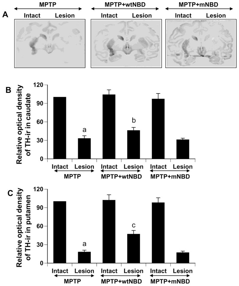
Parkinson's disease (PD) is the most common human neurodegenerative disorder affecting movement, balance, flexibility, and coordination. Despite intense investigation, no effective therapy is available to stop the onset PD or halt its progression. The primate model of PD is considered to be one of the best available models for human PD. Since neuroinflammation plays an important role in the pathogenesis of PD and NF-κB, a proinflammatory transcription factor, participates in the transcription of many proinflammatory molecules, this study evaluates the ability of a peptide corresponding to the NF-κB essential modifier (NEMO)-binding domain (NBD) of IκB kinase (IKK)α or IKKβ to protect dopaminergic neurons in hemiparkinsonian monkeys. First, we found that NF-κB was activated within the substantia nigra pars compacta of 1-methyl-4-phenyl-1,2,3,6-tetrahydropyridine (MPTP)-intoxicated hemiparkinsonian monkeys. However, intramuscular injection of wild type NBD (wtNBD) peptide reduced nigral activation of NF-κB and expression of inducible nitric oxide synthase, protected both the nigrostriatal axis and neurotransmitters, and improved motor functions in hemiparkinsonian monkeys. These findings were specific as mutated NBD peptide did not exhibit such effects. These results may help in the translation of NF-κB-based therapy to PD clinics.


Conflict of interest statement

Conflict of interest: The authors declare that they have no conflict of interest.

Figures

Fig. 1
Fig. 1. Activation of classical NF-κB p65:p50 heterodimer in monkey macrophages
Monkey macrophages were isolated from peripheral blood using the Ficoll-Hypaque method. Briefly, peripheral blood mononuclear cells (PBMC) were separated from erythrocytes by density centrifugation on a Ficoll (Fisher) gradient, washed and resuspended in culture media (5 × 10⁶ cells ml−1). Cells were plated in 12-well plates (1 ml/well) and allowed to adhere for 30 min. Non-adherent cells were removed and adherent cells (mostly macrophages) were washed thrice with RPMI-1640. Four h after plating, macrophages were stimulated with human TNF-α (50 ng/ml) and IL-1β (50 ng/ml) for different time periods followed by monitoring DNA-binding activity of NF-κB (A) in nuclear extracts by electrophoretic mobility shift assay (EMSA) using infrared-labaled consensus NF-κB-binding sequence (5′-AGT TGA GGG GAC TTT CCC AGG C-3′ (Licor Biosciences) as described earlier(Jana and Pahan). B) For the supershift assay, nuclear extract suspended in binding buffer was incubated with 1 μg of anti-p65 Ab, anti-p50 Ab or control IgG for 30 min in ice prior to separation on a 6% polyacrylamide gel. C) Cells preincubated with wtNBD or mNBD peptide for 30 min were stimulated with TNF-α for 1 h followed by monitoring DNA-binding activity of NF-κB by EMSA. Results represent three separate experiments.
Fig. 2
Fig. 2. WtNBD, but not mNBD, peptide treatment decreases the induction of NF-κB p65 in the SNpc of hemiparkinsonian monkeys
Naïve female rhesus monkeys received a right intracarotid injection of MPTP. After 7 d of injection, monkeys displaying classical parkinsonian postures received wtNBD and mNBD peptides (1 mg/kg body wt/2d) via i.m. injection. After 30 d of treatment, nigral sections were immunostained for CD11b (red) & p65 (green) (A) and GFAP (green) & p65 (red) (B). DAPI was used to visualize nucleus. NF-κB p65 positive cells were counted in four nigral sections (two images per slide) of each monkey [MPTP, n=4; (MPTP+wtNBD), n=4; (MPTP+mNBD), n=2] in an Olympus IX81 fluorescence microscope using the MicroSuite imaging software (C). ap < 0.0001 vs control; bp < 0.0001 vs MPTP.
Fig. 3
Fig. 3. WtNBD, but not mNBD, peptide treatment decreases the phosphorylation and acetylation of NF-κB p65 in the SNpc of hemiparkinsonian monkeys
Naïve female rhesus monkeys received a right intracarotid injection of MPTP. After 7 d of injection, monkeys displaying classical parkinsonian postures received wtNBD and mNBD peptides (1 mg/kg body wt/2d) via i.m. injection. After 30 d of treatment, nigral sections were immunostained for HLA-DR (red) & phospho-p65 (green) (A) and HLA-DR (red) & acetylated p65 (green) (B). DAPI was used to visualize nucleus. Cells positive for phospho-p65 (C) and acetylated-p65 (D) were counted in four nigral sections (two images per slide) of each monkey [MPTP, n=4; (MPTP+wtNBD), n=4; (MPTP+mNBD), n=2]. ap < 0.0001 vs control; bp < 0.0001 vs MPTP.
Fig. 4
Fig. 4. WtNBD, but not mNBD, peptide treatment decreases the expression of iNOS in vivo in the nigra of hemiparkinsonian monkeys
Naïve female rhesus monkeys received a right intracarotid injection of MPTP. After 7 d of injection, monkeys displaying classical parkinsonian postures received wtNBD and mNBD peptides (1 mg/kg body wt/2d) via i.m. injection. After 30 d of treatment, nigral sections were immunostained for CD11b (red) & iNOS (green) (A) and GFAP (green) & iNOS (red) (B). DAPI was used to visualize nucleus. Cells positive for iNOS were counted in four nigral sections (two images per slide) of each monkey [MPTP, n=4; (MPTP+wtNBD), n=4; (MPTP+mNBD), n=2] in an Olympus IX81 fluorescence microscope using the MicroSuite imaging software (C). ap < 0.0001 vs control; bp < 0.0001 vs MPTP.
Fig. 5
Fig. 5. WtNBD, but not mNBD, peptide treatment decreases the induction of serum proinflammatory cytokines in hemiparkinsonian monkeys
Naïve female rhesus monkeys received a right intracarotid injection of MPTP. After 7 d of injection, monkeys displaying classical parkinsonian postures received wtNBD and mNBD peptides (1 mg/kg body wt/2d) via i.m. injection. After 30 d of treatment, blood was collected and serum concentration of various proinflammatory cytokines was measured by ELISA (A, TNF-α; B, IL-1β; C, IL-6; D, IL-12; E, IL-1α; F, GM-CSF) by a single investigator blinded to the experimental conditions. Data are means ± SEM of triplicate assays from four monkeys per group. ap < 0.001 vs control; bp < 0.05 vs control; cp < 0.001 vs MPTP; dp < 0.05 vs MPTP.
Fig. 6
Fig. 6. WtNBD, but not mNBD, peptide treatment protects nigral dopaminergic neurons in hemiparkinsonian monkeys
Naïve female rhesus monkeys received a right intracarotid injection of MPTP. After 7 d of injection, monkeys displaying classical parkinsonian postures received wtNBD and mNBD peptides (1 mg/kg body wt/2d) via i.m. injection. After 30 d of treatment, nigral sections were immunostained for TH (A, lower magnification; B, higher magnification). C) Estimates of dopaminergic nigral cell number [MPTP, n=4; (MPTP+wtNBD), n=4; (MPTP+mNBD), n=2] were performed bilaterally using an unbiased design-based counting method (optical fractionator, StereoInvestigator; Microbrightfield, Williston, VT). All counting were performed by a single investigator blinded to the experimental conditions. ap < 0.001 vs MPTP-intact; bp < 0.01 vs MPTP-lesion.
Fig. 7
Fig. 7. WtNBD, but not mNBD, peptide treatment protects striatal fibers in hemiparkinsonian monkeys
Naïve female rhesus monkeys received a right intracarotid injection of MPTP. After 7 d of injection, monkeys displaying classical parkinsonian postures received wtNBD and mNBD peptides (1 mg/kg body wt/2d) via i.m. injection. After 30 d of treatment, striatal sections were immunostained for TH (A). Density of TH fibers in caudate (B) and putamen (C) is expressed as relative to intact side of the MPTP group. Analysis of six different striatal sections of each monkey [MPTP, n=4; (MPTP+wtNBD), n=4; (MPTP+mNBD), n=2] was performed by a single investigator blinded to the experimental conditions. ap < 0.0001 vs MPTP-intact; bp < 0.05 vs MPTP-lesion; cp < 0.001 vs MPTP-lesion.
Fig. 8
Fig. 8. WtNBD, but not mNBD, peptide protects striatal dopamine in hemiparkinsonian monkeys
Naïve female rhesus monkeys received a right intracarotid injection of MPTP. After 7 d of injection, monkeys displaying classical parkinsonian postures received wtNBD and mNBD peptides (1 mg/kg body wt/2d) via i.m. injection. After 30 d of treatment, dopamine levels were measured in dorsomedial caudate (A), ventromedial caudate (B), dorsal putamen (C), ventral putamen (D), and nucleus accumbens (E). HPLC quantification of dopamine was performed by a single investigator blinded to the experimental conditions. Data are means ± SEM of triplicate assays from each monkey [MPTP, n=4; (MPTP+wtNBD), n=4; (MPTP+mNBD), n=2]. ap < 0.001 vs MPTP-intact; bp < 0.05 vs MPTP-lesion.

References

    1. Collier TJ, Kanaan NM, Kordower JH. Ageing as a primary risk factor for Parkinson’s disease: evidence from studies of non-human primates. Nat Rev Neurosci. 12:359–366. - PMC - PubMed
    1. Collier TJ, Steece-Collier K, Kordower JH. Primate models of Parkinson’s disease. Exp Neurol. 2003;183:258–262. - PubMed
    1. Dauer W, Przedborski S. Parkinson’s disease: mechanisms and models. Neuron. 2003;39:889–909. - PubMed
    1. Dehmer T, Lindenau J, Haid S, Dichgans J, Schulz JB. Deficiency of inducible nitric oxide synthase protects against MPTP toxicity in vivo. J Neurochem. 2000;74:2213–2216. - PubMed
    1. Emborg ME, Shin P, Roitberg B, Sramek JG, Chu Y, Stebbins GT, Hamilton JS, Suzdak PD, Steiner JP, Kordower JH. Systemic administration of the immunophilin ligand GPI 1046 in MPTP-treated monkeys. Exp Neurol. 2001;168:171–182. - PubMed

Publication types

LinkOut - more resources