. 2012 Jun 1;2012(6):694-701.
doi: 10.1101/pdb.prot069617.

Imaging neocortical neurons through a chronic cranial window

Imaging neocortical neurons through a chronic cranial window

Anthony Holtmaat et al. Cold Spring Harb Protoc. .

Abstract

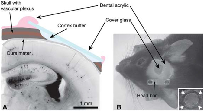
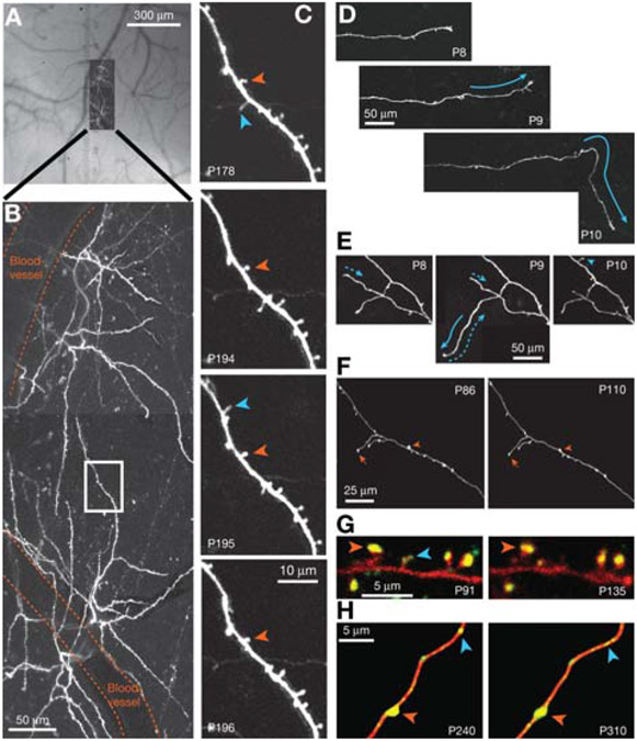
The rich structural dynamics of axonal arbors and neuronal circuitry can only be revealed through direct and repeated observations of the same neuron(s) over time, preferably in vivo. This protocol describes a long-term, high-resolution method for imaging neocortical neurons in vivo, using a combination of two-photon laser scanning microscopy (2PLSM) and a surgically implanted chronic cranial window. The window is used because the skull of most mammals is too opaque to allow high-resolution imaging of cortical neurons. Using this method, it is feasible to image the smallest neuronal structures in the superficial layers of the neocortex, such as dendritic spines and axonal boutons. Because the surface area of the craniotomy is relatively large, this technique is even suitable for use when labeled neurons are relatively uncommon. The surgery and imaging procedures are illustrated with examples from our studies of structural plasticity in the developing or adult mouse brain. The protocol is optimized for adult mice; we have used mice up to postnatal day 511 (P511). With minor modifications, it is possible to image neurons in rats and mice from P2. Most of our studies have used the Thy1 promoter to drive expression of fluorophores in subsets of cortical neurons.


Figures

FIGURE 1.
FIGURE 1.
Experimental preparation for in vivo imaging. (A) Schematic view of the cranial window in cross section superposed on a histologic section of neocortex from a GFP-M transgenic mouse. (B) External view of a cranial window implant; (inset) the circle of thinned bone (arrowheads) around an island of skull (dotted circle) that will be removed during the craniotomy.
FIGURE 2.
FIGURE 2.
Long-term imaging of GFP-expressing pyramidal neurons in transgenic mice. (A) Composite bright-field image of the exposed dura mater and the high-resolution 2PLSM image in B, demonstrating how the vascular tree is used as a reference to locate specific neurons. (B) Top view of dendritic tufts on two pyramidal neurons in layer 5 (projection of a stack of optical sections). Blood vessels and a region of interest (white box) are indicated. (C) Long-term images revealing stable (orange) and transient (blue) dendritic spines from the region of interest (boxed in A). (D,E) Long-term images of postnatal thalamocortical neurons in the developing mouse neocortex showing axonal growth (solid arrows), retraction (dotted arrows), and a new terminaux bouton (blue arrowhead). (Modified, with permission, from Portera-Cailliau et al. 2005.) (F) Examples of stable branches (arrows) and boutons (arrowheads) on thalamocortical axons in the adult mouse neocortex. (Modified, with permission of Elsevier, from De Paola et al. 2006.) (G) Presumptive synaptic contacts indicated by the presence of PSD95-GFP (green) on DsRed-labeled dendritic spines (red); stable (orange arrowhead) and transient (blue arrowhead) synapses are indicated. (H) Synaptophysin-GFP (green) identifies synaptic contacts on axonal boutons expressing DsRed (red). Some synapses are stable (orange arrowhead), but other varicosities appear and disappear (blue arrowheads), possibly indicating synaptic changes. (G and H, Modified, with permission of Macmillan Publishers Ltd., from Holtmaat and Svoboda 2009.) In all images, (P) postnatal day. Panels A–C and F are GFP-M transgenics; D and F are mGFP-L21 transgenics; neurons in F and G were transfected in utero. All images are best projections, as described (Holtmaat et al. 2005; Portera-Cailliau et al. 2005; De Paola et al. 2006).

References

    1. Brown EB, Campbell RB, Tsuzuki Y, Xu L, Carmeliet P, Fukumura D, Jain RK. 2001. In vivo measurement of gene expression, angiogenesis and physiological function in tumors using multiphoton laser scanning microscopy. Nat Med 7: 864–868. - PubMed
    1. Caroni P 1997. Overexpression of growth-associated proteins in the neurons of adult transgenic mice. J Neurosci Methods 71: 3–9. - PubMed
    1. Christie RH, Bacskai BJ, Zipfel WR, Williams RM, Kajdasz ST, Webb WW, Hyman BT. 2001. Growth arrest of individual senile plaques in a model of Alzheimer’s disease observed by in vivo multiphoton microscopy. J Neurosci 21: 858–864. - PMC - PubMed
    1. Denk W, Strickler JH, Webb WW. 1990. Two-photon laser scanning microscopy. Science 248: 73–76. - PubMed
    1. De Paola V, Arber S, Caroni P. 2003. AMPA receptors regulate dynamic equilibrium of presynaptic terminals in mature hippocampal networks. Nat Neurosci 6: 491–500. - PubMed

Publication types

LinkOut - more resources