. 2012 Jul 3;53(8):4458-67.
doi: 10.1167/iovs.11-9166.

Retinal phenotypes in patients homozygous for the G1961E mutation in the ABCA4 gene

Affiliations

Retinal phenotypes in patients homozygous for the G1961E mutation in the ABCA4 gene

Tomas R Burke et al. Invest Ophthalmol Vis Sci. .

Abstract

Purpose: We evaluated the pathogenicity of the G1961E mutation in the ABCA4 gene, and present the range of retinal phenotypes associated with this mutation in homozygosity in a patient cohort with ABCA4-associated phenotypes.

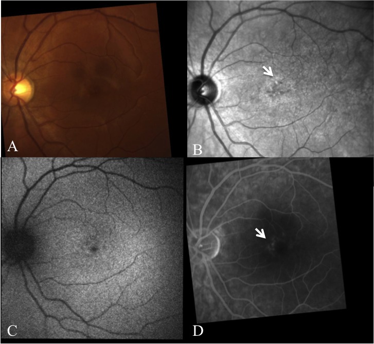
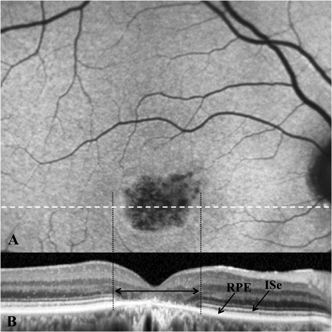
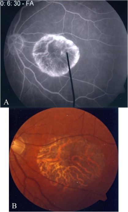
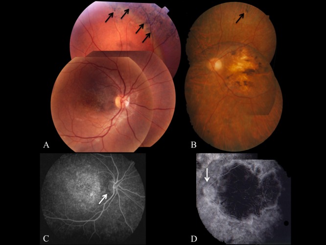
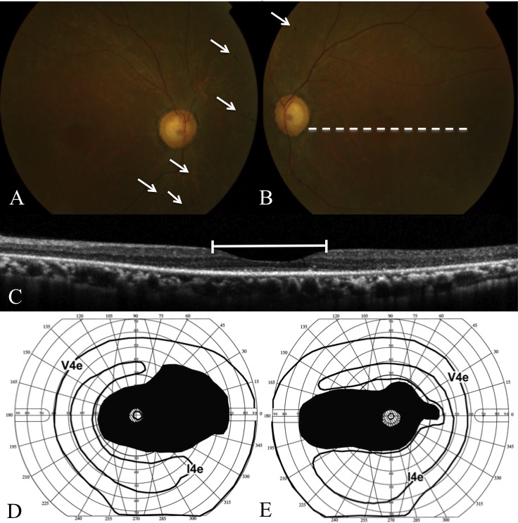
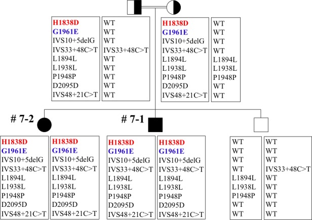
Methods: Patients were enrolled from the ABCA4 disease database at Columbia University or by inquiry from collaborating physicians. Only patients homozygous for the G1961E mutation were enrolled. The entire ABCA4 gene open reading frame, including all exons and flanking intronic sequences, was sequenced in all patients. Phenotype data were obtained from clinical history and examination, fundus photography, infrared imaging, fundus autofluorescence, fluorescein angiography, and spectral domain-optical coherence tomography. Additional functional data were obtained using the full-field electroretinogram, and static or kinetic perimetry.

Results: We evaluated 12 patients homozygous for the G1961E mutation. All patients had evidence of retinal pathology consistent with the range of phenotypes observed in ABCA4 disease. The latest age of onset was recorded at 64 years, in a patient diagnosed initially with age-related macular degeneration (AMD). Of 6 patients in whom severe structural (with/without functional) fundus changes were detected, 5 had additional, heterozygous or homozygous, variants detected in the ABCA4 gene.

Conclusions: Homozygous G1961E mutation in ABCA4 results in a range of retinal pathology. The phenotype usually is at the milder end of the disease spectrum, with severe phenotypes linked to the presence of additional ABCA4 variants. Our report also highlights that milder, late-onset Stargardt disease may be confused with AMD.


Conflict of interest statement

Disclosure: T.R. Burke, None; G.A. Fishman, None; J. Zernant, None; C. Shubert, None; S.H. Tsang, None; R.T. Smith, None; R. Ayyagari, None; R.K. Koenekoop, None; A. Umfress, None; M.L. Ciccarelli, None; A. Baldi, None; A. Iannaccone, None; F.P.M. Cremers, None; C.C.W. Klaver, None; R. Allikmets, None

Figures

Figure 1.
Figure 1.
Fundus photographs (A, B) with corresponding FA images (C, D) of 2 patients (7-2 and 8-1, respectively) with severe STGD1 phenotypes. Note the severe central macular atrophy in both patients, most obvious on FA. While both patients had evidence of peripheral intraretinal pigmentation on fundus photography (black arrows), these were more obvious in patient 7-2. Given the extensive atrophy of the RPE in both patients, detection of the dark choroid sign was not possible. However, partial dark choroid rings were observed in the peripapillary regions (C, D; white arrows).
Figure 2.
Figure 2.
FAF and SD-OCT of the right eye of patient 5. Simultaneous FAF (A) and SD-OCT (B) images were acquired. Note the central macular FAF abnormalities with alternating hypo/hyperautofluorescence surrounded by a perimeter of relative hyperautofluorescence. This corresponded with abnormalities on SD-OCT with absence of the ISe and thinning and irregularity of the RPE (double headed black arrow). Note also the absence of focal hyperautofluorescence, suggestive of flecks, on FAF.
Figure 3.
Figure 3.
FA (with fixation target) and fundus photograph of the left eye of patient 6. The FA (A) showed absence of the dark choroid, as well as evidence of choroidal atrophy (relative hypofluorescence) in the central macula within the region of GA. There was persistence of choroidal perfusion immediately inside the boundary of the region of GA, as evidenced by the irregular ovoid ring of hyperfluorescence in the macula. There was no evidence of flecks or drusen on FA or fundus photography (B).
Figure 4.
Figure 4.
Fundus photographs (A, B) with corresponding FA images (C, D) of 2 patients (7-2 and 8-1, respectively) with severe STGD1 phenotypes. Note the severe central macular atrophy in both patients, most obvious on FA. While both patients had evidence of peripheral intraretinal pigmentation on fundus photography (black arrows), these were more obvious in patient 7-2. Given the extensive atrophy of the RPE in both patients, detection of the dark choroid sign was not possible. However, partial dark choroid rings were observed in the peripapillary regions (C, D; white arrows).
Figure 5.
Figure 5.
Fundus photographs, SD-OCT, and GVF results for patient 10 with a diagnosis of RP. Fundus photographs of the right (A) and left (B) eye are presented. Note the optic disc pallor bilaterally with large round excavated cups. There also was arteriolar narrowing bilaterally with retinal pigment epithelial atrophy along the vascular arcades. A grayish sheen was noted in the mid-periphery bilaterally with areas of intraretinal pigmentation (white arrowheads). (C) SD-OCT image of the left eye corresponding with the dashed line in image (B). The white line indicates the horizontal extent of the central macular region with relative preservation of the inner segment ellipsoid band of the photoreceptors and the outer nuclear layer. Outside this region the photoreceptors, as well as the inner retina, are markedly atrophic. (D, E) GVF of the right and left eye, respectively. Visual function in the central 5° was intact. Absolute ring scotomata were present in the mid-periphery at eccentricities from 5° up to 70° bilaterally. Visual function in the far periphery remained intact.
Figure 6.
Figure 6.
Pedigree for family 7. Patients 7-1 and 7-2 were offspring from a consanguineous marriage. The haplotype for the G1961E mutation, together with the H1838D mutation in this case, is demonstrated clearly. All other variants on the disease haplotype, including the IVS10+5delG, have no effect on splicing or are deemed pathogenic by any other criteria, that is they are frequent (>5%) in the general population and they do not segregate with the disease without the G1961E mutation.

Comment in

References

    1. Blacharski P. Fundus flavimaculatus. : Newsome DA, Retinal Dystrophies and Degenerations. New York, NY: Raven Press; 1988:382
    1. Allikmets R, Singh N, Sun H, et al. A photoreceptor cell-specific ATP-binding transporter gene (ABCR) is mutated in recessive Stargardt macular dystrophy. Nature Genetics. 1997;15:236–246 - PubMed
    1. Jaakson K, Zernant J, Külm M, et al. Genotyping microarray (gene chip) for the ABCR (ABCA4) gene. Hum Mutat. 2003;22:395–403 - PubMed
    1. Yatsenko AN, Shroyer NF, Lewis RA, Lupski JR. Late-onset Stargardt disease is associated with missense mutations that map outside known functional regions of ABCR (ABCA4). Hum Genet. 2001;108:346–355 - PubMed
    1. Maugeri A, van Driel MA, van de Pol DJ, et al. The 2588G-->C mutation in the ABCR gene is a mild frequent founder mutation in the Western European population and allows the classification of ABCR mutations in patients with Stargardt disease. Am J Hum Genet. 1999;64:1024–1035 - PMC - PubMed

Publication types

MeSH terms

Substances