. 2012 Sep 1;21(17):3825-34.
doi: 10.1093/hmg/dds211. Epub 2012 Jun 1.

Evidence for premature aging due to oxidative stress in iPSCs from Cockayne syndrome

Affiliations

Evidence for premature aging due to oxidative stress in iPSCs from Cockayne syndrome

Luciana Nogueira de Sousa Andrade et al. Hum Mol Genet. .

Abstract

Cockayne syndrome (CS) is a human premature aging disorder associated with neurological and developmental abnormalities, caused by mutations mainly in the CS group B gene (ERCC6). At the molecular level, CS is characterized by a deficiency in the transcription-couple DNA repair pathway. To understand the role of this molecular pathway in a pluripotent cell and the impact of CSB mutation during human cellular development, we generated induced pluripotent stem cells (iPSCs) from CSB skin fibroblasts (CSB-iPSC). Here, we showed that the lack of functional CSB does not represent a barrier to genetic reprogramming. However, iPSCs derived from CSB patient's fibroblasts exhibited elevated cell death rate and higher reactive oxygen species (ROS) production. Moreover, these cellular phenotypes were accompanied by an up-regulation of TXNIP and TP53 transcriptional expression. Our findings suggest that CSB modulates cell viability in pluripotent stem cells, regulating the expression of TP53 and TXNIP and ROS production.


Figures
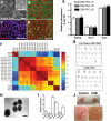
Figure 1.
Figure 1.
Generation and characterization of CSB-iPSCs. (A) Phase contrast image of emerging colonies of CSB-iPSC cells on MEFs 10 days after viral infection. Inset: high magnification of a CSB-iPSC colony displaying cells with a high nucleus/cytoplasm ratio. Bar = 50 μm. (BD) Immunofluorescence staining of pluripotency-associated markers Lin28, Oct3/4, Nanog, SSEA-4 and Sox2 in CSB-iPSC cells. Bar = 50 μm. (E) Nanog, Oct3/4 and Sox2 expression levels in three distinct CSB-iPSC-derived clones by qPCR (n = 3 for each clone). The data are presented as means ± SD. (F) Correlation distances between samples were calculated using vectors of RMA intensity values for all genes on the microarray (MATLAB 7.7, corrcoef). All iPSC clones correlate well with two hESC lines, and less well to donor fibroblasts, indicating reprogrammed iPSCs have similar transcription profiles to hESCs. (G) Absence of gross chromosomal abnormalities on the karyotype of CSB-iPSCs. (H) Differentiation of CSB-iPSCs into embryoid bodies (EB) in vitro. Bar = 50 μm. (I) RT–PCR of relative quantification of endo (AFP and GATA 4), meso (ACT1 and ACTB) and ectodermal (Pax 6 and Nestin) specific genes. The y-axis represents fold change relative to CSB-iPSC expression levels. The data are presented as means ± SD (n = 3 for each clone). (J) Teratoma obtained from Control  and CSB-iPSCs. The detail reveals the lack of blood vessels in CSB-iPSC derived teratomas. Bar = 100 μm.
Figure 2.
Figure 2.
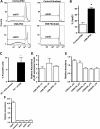
Cell death in CSB-iPSC. (A and B) Flow cytometry analysis of dead cells (represented by sub-G1 content) in CSB-iPSCs and fibroblasts. Each bar represents the mean ± SD (n = 2–7). Statistical analyses were performed using t-test one-tailed and differences were considered significant for P < 0.05 (indicated by asterisk). Flow data from representative samples are shown. In (C), cell death analysis of human H9 ES transfected with a control (shRNA-Scrambled) or plasmid expressing shRNA against CSB (shRNA-CSB). Each bar represents the mean ± SD (n = 3) of H9 ES dead cells. The y-axis represents the percentage of propidium iodide (PI) positive cells (dead cells) determined by flow cytometry. Statistical analyses were performed using t-test one-tailed and differences were considered significant for P < 0.05 (indicated by asterisk). (D) RT–PCR for TP53 expression in distinct clones of both control and CSB-iPSCs. The y-axis represents fold change relative to TP53 levels in Control iPSCs. The data are presented as mean (n = 3). (E) Cell cycle regulators mRNA levels in CSB-iPSCs. The y-axis represents fold change relative to each gene level in control. The data are presented as means ± SD (triplicate for each clone). (F) mRNA level measurements by microarray of cell mediators of oxidative stress in CSB-iPSCs. The y-axis represents fold change relative to each gene level in control. The data are presented as means ± SD (triplicate for each clone).
Figure 3.
Figure 3.
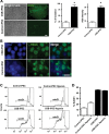
Quantification of the production of ROS in control and CSB-iPSCs. (A) Images were taken after incubation with H2DCFDA in control and CSB-iPSCs plated at the same density (bar = 50 μm). Bar graphs in the right show percentage of cells producing ROS and fluorescence intensity. Mean ± SD (n = 3). Statistical analyses were performed using t-test one-tailed and differences were considered significant for P < 0.05 (indicated by asterisk). (B) DDR activation in CSB-iPSCs. Immunofluorescence staining shows the presence of γ-H2AX at similar levels in both CSB and Control iPSCs. Representative images are shown. Bar = 15 μm. (C and D) Cell death evaluation under hypoxia. Flow cytometry analysis of sub-G1 content in control and CSB-iPSCs cultivated under hypoxia conditions for 24h. Bar graph shows the percentage of dead cells in normoxia and hypoxia. Representative samples are shown.
Figure 4.
Figure 4.
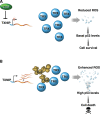
CSB plays a role in pluripotent stem cell survival. According to our model, CSB inhibits TXNIP transcription, preventing the blockage of TRX scavenger activity and favoring cell survival (A). In contrast, the absence of functional CSB would increase TXNIP transcription and its interaction with TRX, leading to an increase in intracellular ROS levels. As a consequence, TP53 is up-regulated, triggering cell death (B).

References

    1. Weidenheim K.M., Dickson D.W., Rapin I. Neuropathology of Cockayne syndrome: evidence for impaired development, premature aging, and neurodegeneration. Mech. Ageing Dev. 2009;130:619–636. - PubMed
    1. Laugel V., Dalloz C., Durand M., Sauvanaud F., Kristensen U., Vincent M.C., Pasquier L., Odent S., Cormier-Daire V., Gener B., et al. Molecular analysis of mutations in the CSB (ERCC6) gene in patients with Cockayne syndrome. Mutation update for the CSB/ERCC6 and CSA/ERCC8 genes involved in Cockayne syndrome. Hum. Mutat. 2010;31:113–126. - PubMed
    1. Citterio E., Van Den Boom V., Schnitzler G., Kanaar R., Bonte E., Kingston R.E., Hoeijmakers J.H., Vermeulen W. ATP-dependent chromatin remodeling by the Cockayne syndrome B DNA repair-transcription-coupling factor. Mol. Cell Biol. 2000;20:7643–7653. - PMC - PubMed
    1. Newman J.C., Bailey A.D., Weiner A.M. Cockayne syndrome group B protein (CSB) plays a general role in chromatin maintenance and remodeling. Proc. Natl Acad. Sci. USA. 2006;103:9613–9618. - PMC - PubMed
    1. Licht C.L., Stevnsner T., Bohr V.A. Cockayne syndrome group B cellular and biochemical functions. Am. J. Hum. Genet. 2003;73:1217–1239. - PMC - PubMed

Publication types

Substances