. 2012 Sep;194(17):4494-504.
doi: 10.1128/JB.00011-12. Epub 2012 Jun 1.

Daptomycin-mediated reorganization of membrane architecture causes mislocalization of essential cell division proteins

Affiliations

Daptomycin-mediated reorganization of membrane architecture causes mislocalization of essential cell division proteins

Joe Pogliano et al. J Bacteriol. 2012 Sep.

Abstract

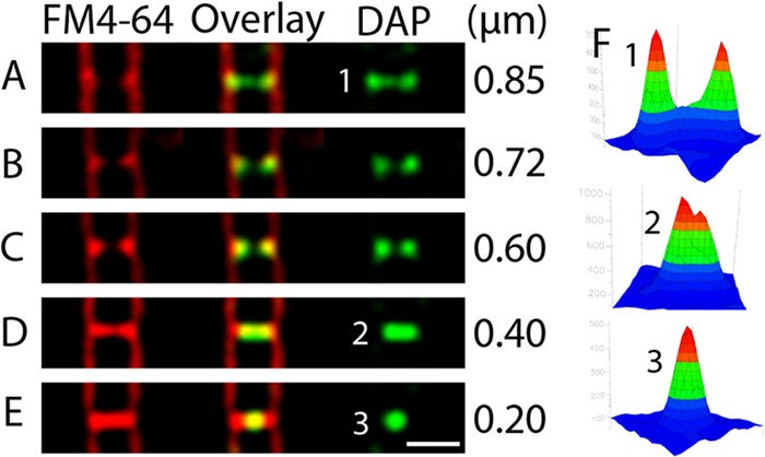
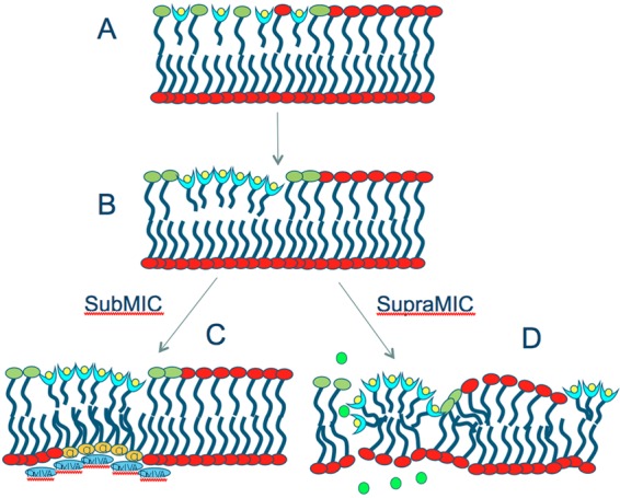
Daptomycin is a lipopeptide antibiotic used clinically for the treatment of certain types of Gram-positive infections, including those caused by methicillin-resistant Staphylococcus aureus (MRSA). Details of the mechanism of action of daptomycin continue to be elucidated, particularly the question of whether daptomycin acts on the cell membrane, the cell wall, or both. Here, we use fluorescence microscopy to directly visualize the interaction of daptomycin with the model Gram-positive bacterium Bacillus subtilis. We show that the first observable cellular effects are the formation of membrane distortions (patches of membrane) that precede cell death by more than 30 min. Membrane patches are able to recruit the essential cell division protein DivIVA. Recruitment of DivIVA correlates with membrane defects and changes in cell morphology, suggesting a localized alteration in the activity of enzymes involved in cell wall synthesis that could account for previously described effects of daptomycin on cell wall morphology and septation. Membrane defects colocalize with fluorescently labeled daptomycin, DivIVA, and fluorescent reporters of peptidoglycan biogenesis (Bocillin FL and BODIPY FL-vancomycin), suggesting that daptomycin plays a direct role in these events. Our results support a mechanism for daptomycin with a primary effect on cell membranes that in turn redirects the localization of proteins involved in cell division and cell wall synthesis, causing dramatic cell wall and membrane defects, which may ultimately lead to a breach in the cell membrane and cell death. These results help resolve the longstanding questions regarding the mechanism of action of this important class of antibiotics.


Figures

Fig 1
Fig 1
Microscopic evaluation of the cell biological effects of daptomycin exposure. (A) Growth curve of cells in different concentrations of daptomycin demonstrating that cells grown in 1 μg/ml, 2 μg/ml, or 3 μg/ml daptomycin have growth rates identical to those of cells grown without antibiotic. In contrast, cells grown with 10 μg/ml daptomycin fail to grow and lyse after approximately 2.5 h. Note that B. subtilis is prone to lysis due to the activation of autolysins, whereas S. aureus does not exhibit lysis in the presence of daptomycin. (B) The percentage of cells containing bright sytox green staining increases over time when the cells grow in a lethal concentration of daptomycin (10 μg/ml). The cells displaying bright sytox green staining were counted and binned into groups, and the percentages of cells displaying bright sytox green staining were plotted. Zero to 10 min, 0% sytox green-positive cells, n = 146; 11 to 20 min, 7% sytox green-positive cells, n = 300; 21 to 30 min, 11% sytox green-positive cells, n = 314; 31 to 45 min, 74% sytox green-positive cells, n = 57; 46 to 60 min, 94% sytox green-positive cells, n = 446. (C to H) Time course of cells grown on an agarose pad containing FM 4-64, sytox green, and a lethal dose of daptomycin (10 μg/ml) demonstrating that cells suffer membrane deformations and ultimately lose their membrane integrity and stain brightly with sytox green. The cells in panels C and D have normal membrane and sytox green staining. The cells in panels E to G have membrane defects but normal (low) sytox green staining. The cells in panel H have high sytox green staining. (I to L) Time lapse microscopy demonstrating that membrane deformations precede cell lysis. (I) After 25 min of growth in the presence of daptomycin, a cell contains a large membrane protrusion (I) but displays low sytox green staining (K). (J and L) Five minutes later, the membrane has become permeable and the cell stains brightly with sytox green. The top images in panels I and J show membranes only. The bottom images show membranes and sytox green. (K) Quantification of sytox fluorescence intensity for the cell in panel I. (L) Quantification of sytox fluorescence intensity for the cell in panel J.
Fig 2
Fig 2
Daptomycin-BODIPY staining of B. subtilis during growth. Cells were labeled for 10 min with 16 μg/ml daptomycin-BODIPY (green) and stained with FM 4-64 (red) during vegetative growth. All panels are at the same scale (bar, 1 μm). Daptomycin labels the sidewalls as discrete foci and intensely stains active division sites. (A to C) A single cell was labeled with FM 4-64 (A), daptomycin-BODIPY (C), or both (B). (D and E) A single cell showing that medial (D) and top (E) focal planes reveal different foci surrounding the cell. (F) Three-dimensional (3D) intensity plot corresponding to the region boxed in panel E showing that septal staining (large peaks) is more intense than sidewall staining. (G) Middle focal plane of a chain of cells labeled with daptomycin-BODIPY (green). (H and I) A single nascent septum labeled with daptomycin-BODIPY (green). The boxed region was reconstructed in three dimensions and then rotated through 90° (panel I, from left to right), demonstrating that the daptomycin-BODIPY fluorescence at division sites circumnavigates the cell.
Fig 3
Fig 3
Demonstration that daptomycin-BODIPY (DAP-Bodipy) intensely stains nascent septa but is generally absent from complete septa. Cells were labeled for 10 min with 16 μg/ml daptomycin-BODIPY as described in Materials and Methods. (A to C) Images showing cell membranes stained with FM 4-64 (A), daptomycin-BODIPY (B), or both (C). Scale bar, 1 μm. (D to F) Fluorescence intensity plots of an incomplete septum (1 in panel A, enlarged in panel F) showing that the peaks of membrane fluorescence corresponding to septal invagination (D) coincide with peaks of daptomycin-BODIPY fluorescence (E). Scale bar, 1 μm. (G to I) Fluorescence intensity plots for a complete septum (2 in panel A, enlarged in panel I) showing that the peaks of daptomycin-BODIPY fluorescence (H) do not coincide with the peaks of membrane fluorescence (G).
Fig 4
Fig 4
Five cells containing septa at various stages of constriction. Daptomycin-BODIPY stains nascent septa first as a ring, then as a smaller ring, then as a small band of fluorescence, and finally, when the septum appears to be complete, as a bright focus. The width of the daptomycin-BODIPY (DAP) labeling is provided at the right of each panel. Note that most septa (76%; n = 55) that appear to be complete, as in panel E, have no daptomycin-BODIPY staining. Scale bar, 1 μm. All panels are at the same scale.
Fig 5
Fig 5
Daptomycin-BODIPY localization during sporulation. The arrows indicate the positions of intense labeling. At 1.5 h (A and B) and 2 h (C) after the onset of sporulation, cells were labeled with 16 μg/ml daptomycin-BODIPY (green) and stained with FM 4-64 (red) as described in Materials and Methods. (A) Cells that have not yet synthesized a polar sporulation septum contain two polar daptomycin-BODIPY rings (blue arrows). (B and C) Cells that have completed polar septation and initiated engulfment show intense staining at the leading edge of the engulfing mother cell membrane (white arrows). Scale bar, 1 μm. All panels are at the same scale.
Fig 6
Fig 6
Cells grown in the presence of sublethal concentrations of daptomycin have a striking change in morphology. (A to F) Images of B. subtilis (PY79) after 2 h of growth in the presence of a sublethal concentration of daptomycin (1 μg/ml). The cell membranes are stained red with FM 4-64. (G to J) Daptomycin (Dap)-BODIPY localizes to membrane patches and to the membrane at the interiors of curved regions at sublethal concentrations. The panels show membrane staining (top), daptomycin-BODIPY labeling (bottom), or both (middle). PY79 was grown in LB with the sublethal dose of 10 μg/ml daptomycin-BODIPY for 2 h at 37°C. All panels are at the same scale. Scale bars, 1 μm.
Fig 7
Fig 7
DivIVA-GFP targets positions of membrane rearrangement and bent regions of sub-MIC daptomycin-treated cells, but SpoVM-GFP is specifically excluded. (A) In wild-type cells, DivIVA-GFP localizes to membrane invaginations during and after cell division. The fluorescence intensity graphs for membranes (FM 4-64) and DivIVA-GFP correspond to the boxed region. (B) DivIVA-GFP localizes to membrane patches formed in cells grown in a sublethal concentration of daptomycin (2 μg/ml). The three-dimensional graphs of fluorescence intensity corresponding to the boxed region show that regions containing concentrated membranes recruit DivIVA-GFP. (C and D) Two additional examples of cells grown in sublethal concentrations of daptomycin (2 μg/ml) showing that DivIVA-GFP is localized to the bent regions of the cells (arrows), where membrane patches occur. (E) SpoVM-GFP uniformly decorates the membrane in wild-type cells. Three-dimensional fluorescence intensity graphs of SpoVM-GFP and FM 4-64 are shown for the boxed region. (F and G) SpoVM-GFP is excluded from daptomycin-induced membrane patches. The three-dimensional fluorescence intensity graphs show that SpoVM-GFP is uniformly distributed around the cell and does not accumulate within the membrane patches. All panels are at the same scale. Scale bars, 1 μm.
Fig 8
Fig 8
Fluorescent vancomycin targets positions of active peptidoglycan biogenesis, accumulating within septa in control cells and also in membrane patches and bent regions of sub-MIC daptomycin-treated cells. Cells were stained with the vital membrane stains FM 4-64 (1 μg/ml), DAPI (2 μg/ml), and a fluorescent derivative of vancomycin, as described in Materials and Methods. (A) In cells not treated with daptomycin, Van-BDP (0.5 μg/ml; ∼1× MIC) intensely stains septa and only very faintly stains the sidewalls, as previously shown (9, 17, 54). The septum in the middle of the cell is just beginning to divide and accumulates Van-BDP as a ring of fluorescence, which appears as two dots in the medial focal plan. Van-BDP forms an intense band at completed septa, as previously shown (9, 17, 54). Scale bar, 1 μm. (B) Two examples showing that Van-BDP localizes to the membrane patches and bent regions that are formed in cells grown in a sublethal concentration of daptomycin (2 μg/ml). Scale bar, 1 μm.
Fig 9
Fig 9
The fluorescent penicillin V derivative Bocillin FL targets positions of active peptidoglycan biogenesis, accumulating within septa in control cells and in bent regions of sub-MIC daptomycin-treated cells. Cells were stained with the vital membrane stains FM 4-64 (1 μg/ml), DAPI (2 μg/ml), and Bocillin FL as described in Materials and Methods. (A) In cells not treated with daptomycin, Bocillin FL more intensely stains regions actively undergoing septation, as expected. The cell on the left is just beginning to divide and accumulates fluorescent Bocillin as a ring of fluorescence. The cell on the right has nearly completed cell division, and Bocillin FL accumulates as a small focus in the center of the septum. Scale bar, 1 μm. (B) Bocillin FL intensely stains the membrane patches and bent regions formed in cells grown in a sublethal concentration of daptomycin (2 μg/ml). Two examples are shown. Scale bar, 1 μm.
Fig 10
Fig 10
A refined model for the mechanism of action of daptomycin (light-blue “cups” with yellow circles). See the text for details. Shown are phosphatidylglycerol (green lipids), cardiolipin (yellow lipids), potassium ions (green circles), and DivIVA (blue ovals).

References

    1. Abanes-De Mello A, Sun YL, Aung S, Pogliano K. 2002. A cytoskeleton-like role for the bacterial cell wall during engulfment of the Bacillus subtilis forespore. Genes Dev. 16:3253–3264 - PMC - PubMed
    1. Arias CA, et al. 2011. Genetic basis for in vivo daptomycin resistance in enterococci. N. Engl. J. Med. 365:892–900 - PMC - PubMed
    1. Baltz RH. 2009. Daptomycin: mechanisms of action and resistance, and biosynthetic engineering. Curr. Opin. Chem. Biol. 13:144–151 - PubMed
    1. Bramkamp M, van Baarle S. 2009. Division site selection in rod-shaped bacteria. Curr. Opin. Microbiol. 12:683–688 - PubMed
    1. Broder DH, Pogliano K. 2006. Forespore engulfment mediated by a ratchet-like mechanism. Cell 126:917–928 - PMC - PubMed

Publication types

MeSH terms

LinkOut - more resources