. 2012 Jul 19;120(3):592-602.
doi: 10.1182/blood-2012-02-408732. Epub 2012 Jun 1.

Impaired natural killer cell self-education and "missing-self" responses in Ly49-deficient mice

Affiliations

Impaired natural killer cell self-education and "missing-self" responses in Ly49-deficient mice

Simon Bélanger et al. Blood. .

Erratum in

  • Blood. 2013 Oct 3;122(14):2525

Abstract

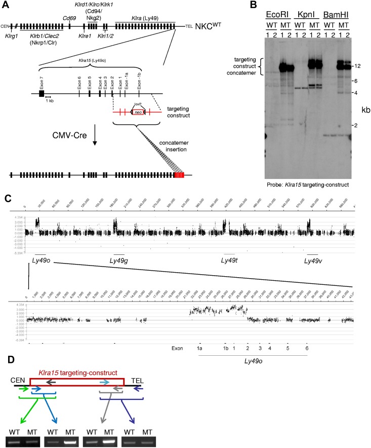
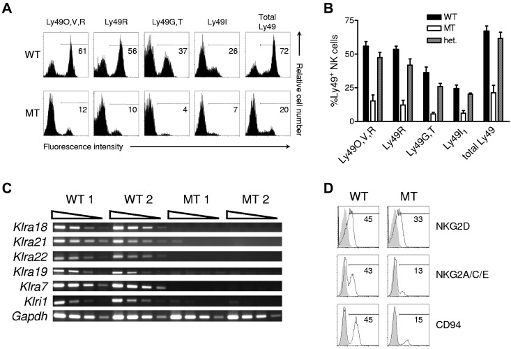
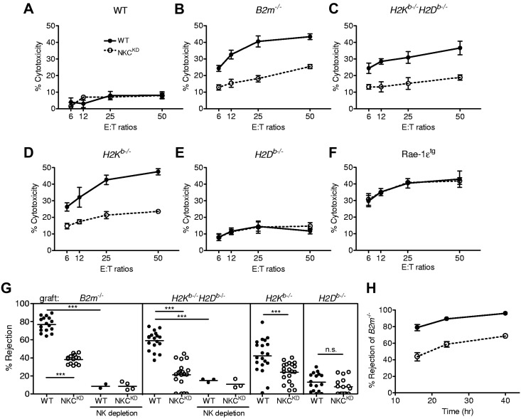
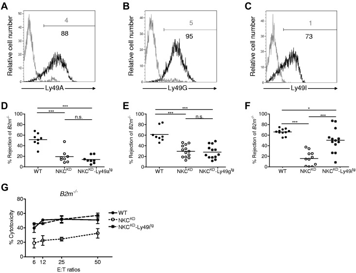
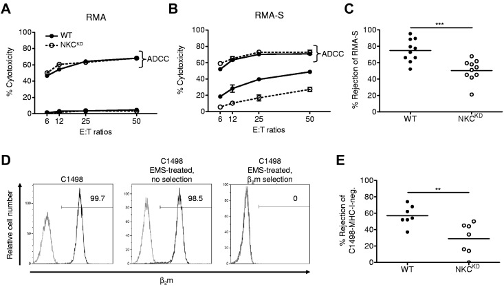
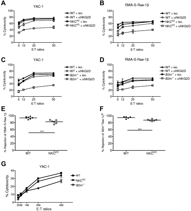
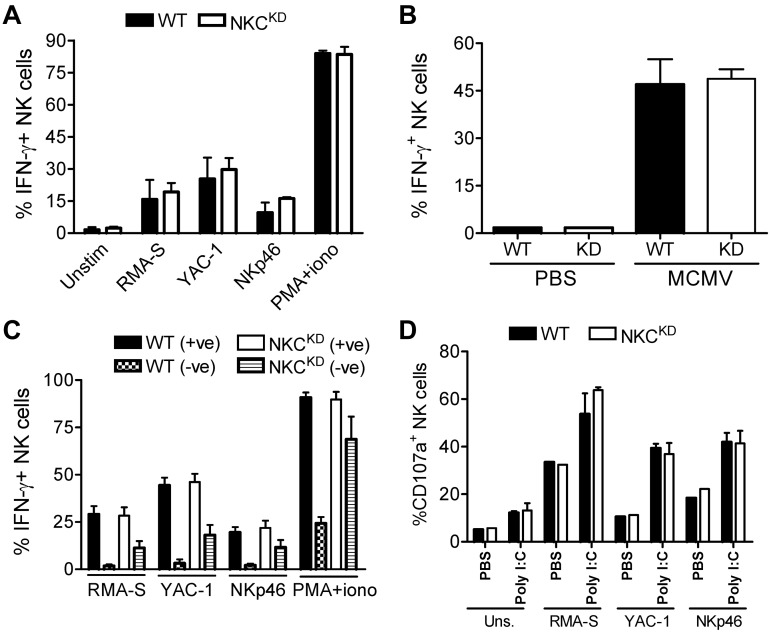
Ly49-mediated recognition of MHC-I molecules on host cells is considered vital for natural killer (NK)-cell regulation and education; however, gene-deficient animal models are lacking because of the difficulty in deleting this large multigene family. Here, we describe NK gene complex knockdown (NKC(KD)) mice that lack expression of Ly49 and related MHC-I receptors on most NK cells. NKC(KD) NK cells exhibit defective killing of MHC-I-deficient, but otherwise normal, target cells, resulting in defective rejection by NKC(KD) mice of transplants from various types of MHC-I-deficient mice. Self-MHC-I immunosurveillance by NK cells in NKC(KD) mice can be rescued by self-MHC-I-specific Ly49 transgenes. Although NKC(KD) mice display defective recognition of MHC-I-deficient tumor cells, resulting in decreased in vivo tumor cell clearance, NKG2D- or antibody-dependent cell-mediated cytotoxicity-induced tumor cell cytotoxicity and cytokine production induced by activation receptors was efficient in Ly49-deficient NK cells, suggesting MHC-I education of NK cells is a single facet regulating their total potential. These results provide direct genetic evidence that Ly49 expression is necessary for NK-cell education to self-MHC-I molecules and that the absence of these receptors leads to loss of MHC-I-dependent "missing-self" immunosurveillance by NK cells.


Figures

Figure 1
Figure 1
The Klra gene cluster of Klra15-targeted (mutant) mice contains a concatemer insertion of the Klra15-targeting construct. (A) A schematic representation of the natural killer gene complex (NKC) and the targeting-construct produced for the Klra15 gene. (B) Southern blot analysis of wild-type (WT) and mutant thymus DNA digested with EcoRI, KpnI, or BamHI and probed with the Klra15-targeting construct depicted in panel A. (C) Array comparative genomic hybridization profile of the Klra cluster in mutant mice with a blow-up of the Klra15 gene (below). The x-axis represents the genomic region tiled on the microarrays, and the y-axis Shows differences in copy number between WT and mutant genomic DNA (log2 ratio [MT/WT]). Positive values indicate regions showing copy number increases in the NKC knockdown (NKCKD) genome. (D) PCR analysis of the Klra15-targeting construct copy number in mutant genomic DNA. Data are representative of ≥ 3 similar experiments, except for array comparative genomic hybridization analysis, which was performed twice. These experiments were performed with mice on the 129S1 background.
Figure 2
Figure 2
Klra15-targeted mice show a drastic reduction in telomeric NKC gene expression. (A) NK cells (DX5+TCRβ) from the spleens of WT or mutant mice were analyzed for Ly49 expression with mAbs 4E5 (which binds Ly49O/V/R in mice with a 129-strain Klra gene cluster), 12A8 (Ly49R), 4D11 (Ly49G/T), and 14B11 (Ly49I) individually or as a cocktail (Total Ly49). The percentage of NK cells positively stained is indicated. (B) The mean ± SD percentage of different splenic NK-cell subsets present in WT, mutant, and heterozygous littermates (n = 5) is shown graphically. (C) Semiquantitative RT-PCR of cDNA for the indicated genes was performed on total RNA obtained from lymphokine-activated killer cells prepared from 2 individual WT or mutant mouse spleens. (D) The expression of NKG2/CD94 family of receptors on splenic NK cells from WT or mutant littermates. Data are representative of ≥ 3 similar experiments. These experiments were performed with mice on the 129S1 background.
Figure 3
Figure 3
Natural killer gene complex knockdown (NKCKD) NK cells exhibit defective in vitro killing of MHC-I–deficient ConA blasts, and NKCKD mice exhibit reduced rejection of MHC-I–deficient splenocytes in vivo. The ability of lymphokine-activated killer (LAK) cells from WT and NKCKD to kill splenic ConA blast target cells from (A) B6 (WT), (B) B2m−/−, (C) H2Kb−/−H2Db−/−, (D) H2Kb−/−, (E) H2Db−/−, and (F) Rae-1ϵ transgenic (Rae-1ϵtg) mice was tested by 51Cr-release assay. Data are represented as the mean ± SD percentage of chromium release from triplicate wells. Data are representative of 3 similar experiments. Cytotoxicity experiments were performed with mice on the B6 background. (G) The ability of WT and NKCKD mice to reject CFSE-labeled splenocytes from B2m−/−, H2Kb−/−H2Db−/−, H2Kb−/−, and H2Db−/− mice was tested by flow cytometry of recipient splenocytes 16 hours after injection. Each symbol represents the data from an individual mouse, and the small horizontal lines indicate the mean. Some mice of each strain were pretreated with anti–asialo-GM1 Ab to deplete NK cells before injection of CFSE-labeled cells. Data are pooled from 3 to 5 independent experiments. Groups differed significantly as shown (***P < .001; NS, not significant). (H) Time-course rejection assay of B2m−/− splenocytes by WT and NKCKD mice. Data are displayed as the mean ± SEM rejection of 6 individual mice. Splenocyte rejection experiments were performed with 129S1 background mice.
Figure 4
Figure 4
Loss of MHC-I-immunosurveillance in NKCKD mice is because of silencing of Ly49 that bind to self–MHC-I. Three different Ly49-transgenes were introduced into NKCKD mice by breeding. (A-C) Flow cytometric analysis of Ly49 expression in Ly49tg-NKCKD NK cells. Ly49 staining of NK cells is shown for (A) NKCKD-Ly49Atg, (B) NKCKD-Ly49Gtg, and (C) NKCKD-Ly49Itg mice as a black histogram. The gray histogram shows control staining of splenic NK cells from NKCKDmice. (D-F) Specific rejection of B2m−/− splenocytes by (D) Ly49Atg-NKCKD mice, (E) Ly49Gtg-NKCKD mice, (F) Ly49Itg-NKCKD mice in comparison to WT or NKCKD mice. Each symbol represents the data from a single mouse. Data are pooled from 3 to 5 independent experiments. Groups differed significantly as shown (*P < .05; ***P < .001; NS indicates not significant). (G) LAK prepared from WT, NKCKD, and NKCKD-Ly49itg mice were used as effector cells in a 51Cr-release assay against B2m−/− ConA blasts. These experiments were performed with mice on the B6 background.
Figure 5
Figure 5
NKCKD NK cells exhibit reduced cytotoxicity toward MHC-I–deficient tumor cells in vitro and in vivo. (A-B) The ability of WT versus NKCKD lymphokine-activated killer (LAK) cells to kill tumor target cells was tested by 51Cr-release assay. LAK cells generated from WT and NKCKD mice were used as effectors cells against untreated (A) RMA, (B) RMA-S target cells, or targets precoated with anti-Thy1.2 mAb to test antibody-dependent cellular cytotoxicity (ADCC) function. The data are displayed as the mean ± SD percentage of chromium release from triplicate wells. (C) In vivo rejection of RMA-S relative to RMA cells was assessed. The mean rejection is indicated by a horizontal line. Each symbol represents the data from a single mouse. (D) Production of a MHC-I–negative C1498 subline. Flow cytometry for β2m (dark histogram) is shown for the indicated C1498 lines. The gray histogram indicates isotype control mAb staining. The percentage of cells positively staining for β2m is indicated. (E) In vivo rejection of C1498-MHC-I–negative cells versus C1498-MHC-I–positive tumor cells. Note that both cell lines received EMS treatment. Data are representative of ≥ 3 similar experiments. Groups differed significantly as shown (**P < .01; ***P < .001). These experiments were performed with mice on the B6 background.
Figure 6
Figure 6
Activation via NKG2D overcomes deficient missing-self responses by natural killer gene complex knockdown (NKCKD) and B2m−/− NK cells to tumor cells. (A-B) The cytotoxic potential of WT versus NKCKD LAK cells against (A) YAC-1 and (B) RMA-S-Rae-1β target cells was assessed by 51Cr-release assay in the presence or absence of blocking anti-NKG2D. (C-D) Similarly, cytotoxicity of WT versus B2m−/− LAK cells was assessed against (C) YAC-1 and (D) RMA-S-Rae-1β target cells in the presence or absence of blocking anti-NKG2D. The data are displayed as the mean ± SD percentage of chromium release from triplicate wells. (E-F) In vivo rejection of (E) RMA-S-Rae-1β relative to RMA cells or of (F) B2m−/−Rae-1etg splenocytes relative to WT splenocytes was assessed. The mean rejection is indicated by a horizontal line. Each symbol represents the data from a single mouse. Data are representative of ≥ 3 similar experiments. Groups differed significantly as shown (***P < .001). (G) Splenocytes from the indicated mouse strains were used in cytotoxicity assays against 51Cr-labeled YAC-1 cells at the indicated effector-to-target ratios. The data are displayed as the mean ± SD of chromium release from triplicate wells. These experiments were performed with mice on the B6 background.
Figure 7
Figure 7
NKCKD NK cells display normal IFN-γ production and degranulation responses. (A) Splenocytes from WT or NKCKD mice pretreated with poly(I:C) were incubated with the indicated tumor cells, on plates coated with anti-NKp46 mAb or with phorbol 12-myristate 13-acetate (PMA)/ionomycin. After 5 hours intracellular staining was performed to assess IFN-γ production by flow cytometry. (B) The frequency of IFN-γ+ NK cells was assessed in the spleens of the indicated mouse strains after 36 hours of infection with Smith strain murine CMV (MCMV). (C) The same assay as in panel A was performed, but splenocytes were additionally stained with anti-Ly49 and NKG2A mAb. Data are shown as the percentage of IFN-γ+ cells among Ly49/NKG2+ and Ly49/NKG2 subsets. (D) CD107a levels on the surface of NK cells were evaluated by flow cytometry after the indicated tumor or mAb stimulations. Mice were pretreated with poly(I:C) or with a PBS control. Data are representative of ≥ 3 similar experiments, except for degranulation assays which were performed twice. Three individual mice were used as a source of splenocytes for each experiment (n = 3). These experiments were performed with mice on the B6 background.

References

    1. Biron CA, Byron KS, Sullivan JL. Severe herpesvirus infections in an adolescent without natural killer cells. N Engl J Med. 1989;320(26):1731–1735. - PubMed
    1. Orange JS. Human natural killer cell deficiencies. Curr Opin Allergy Clin Immunol. 2006;6(6):399–409. - PubMed
    1. Kärre K, Ljunggren HG, Piontek G, Kiessling R. Selective rejection of H-2-deficient lymphoma variants suggests alternative immune defence strategy. Nature. 1986;319(6055):675–678. - PubMed
    1. Carlyle JR, Mesci A, Fine JH, et al. Evolution of the Ly49 and Nkrp1 recognition systems. Semin Immunol. 2008;20(6):321–330. - PubMed
    1. Makrigiannis AP, Patel D, Goulet ML, Dewar K, Anderson SK. Direct sequence comparison of two divergent class I MHC natural killer cell receptor haplotypes. Genes Immun. 2005;6(2):71–83. - PubMed

Publication types

MeSH terms