. 2012 Jul 19;120(3):636-46.
doi: 10.1182/blood-2012-04-424143. Epub 2012 Jun 1.

Tissue factor promotes activation of coagulation and inflammation in a mouse model of sickle cell disease

Affiliations

Tissue factor promotes activation of coagulation and inflammation in a mouse model of sickle cell disease

Pichika Chantrathammachart et al. Blood. .

Abstract

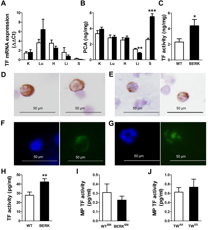
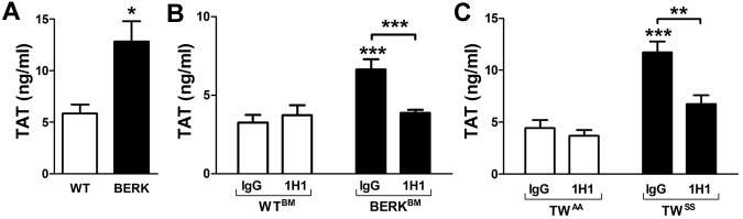
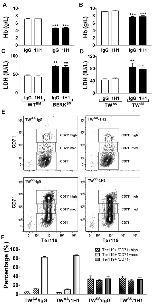
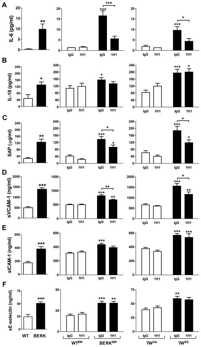
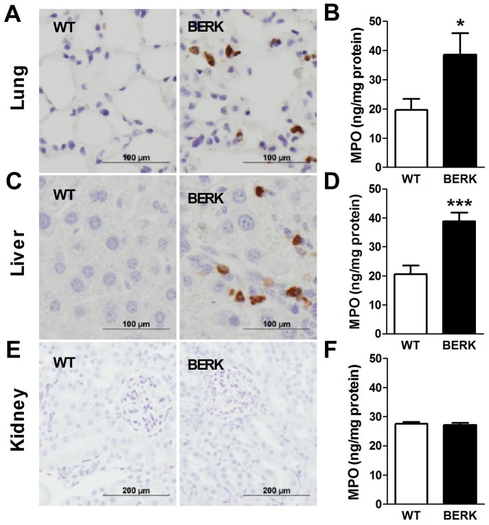
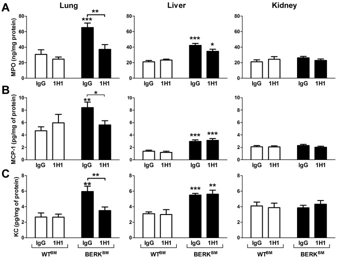
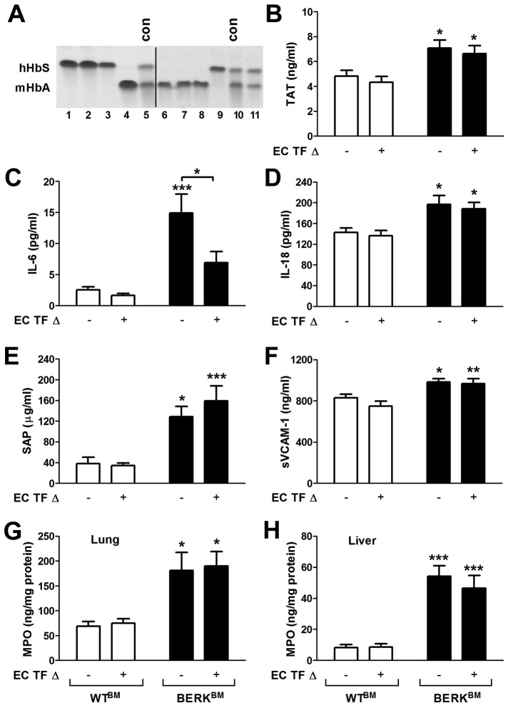
Sickle cell disease (SCD) is associated with a complex vascular pathophysiology that includes activation of coagulation and inflammation. However, the crosstalk between these 2 systems in SCD has not been investigated. Here, we examined the role of tissue factor (TF) in the activation of coagulation and inflammation in 2 different mouse models of SCD (BERK and Townes). Leukocytes isolated from BERK mice expressed TF protein and had increased TF activity compared with control mice. We found that an inhibitory anti-TF antibody abrogated the activation of coagulation but had no effect on hemolysis or anemia. Importantly, inhibition of TF also attenuated inflammation and endothelial cell injury as demonstrated by reduced plasma levels of IL-6, serum amyloid P, and soluble vascular cell adhesion molecule-1. In addition, we found decreased levels of the chemokines MCP-1 and KC, as well as myeloperoxidase in the lungs of sickle cell mice treated with the anti-TF antibody. Finally, we found that endothelial cell-specific deletion of TF had no effect on coagulation but selectively attenuated plasma levels of IL-6. Our data indicate that different cellular sources of TF contribute to activation of coagulation, vascular inflammation, and endothelial cell injury. Furthermore, it appears that TF contributes to these processes without affecting intravascular hemolysis.


Figures

Figure 1
Figure 1
TF expression in sickle cell mice. (A) TF mRNA expression and (B) procoagulant activity (PCA) in organs of WT (open bar; n = 9) and BERK mice (filled bar; n = 8). K indicates kidney; Lu, lung; H, heart; Li, liver; and S, spleen. Asterisks directly above the bars indicate statistically significant difference compared with WT controls: **P < .01 and ***P < .001. (C) TF-dependent PCA activity in the spleens of WT (open bar) and BERK mice (filled bar). *P < .05. Immunocytochemistry (D-E) or immunofluorescence (F-G) staining demonstrates TF-positive leukocytes with nuclear morphology resembling monocytes (D,F) or neutrophils (E,G). TF is shown as brown staining (top panels) or green staining (bottom panels). Nuclei are stained blue. (H) TF activity of the leukocytes isolated from the same volume of blood obtained from WT (n = 13) or BERK (n = 10) mice. **P < .01. (I) TF activity of the microparticles (MP TF) isolated from the plasma of WT (open bar; n = 6) and BERK (filled bar; n = 15) mice. (J) MP TF activity in TWAA (open bar; n = 13) and TWSS (filled bar; n = 17) mice.
Figure 2
Figure 2
Activation of coagulation in sickle cell mice. (A) Plasma TAT levels in WT (open bar; n = 6) and BERK (filled bar; n = 16) mice (both on a similar mixed genetic background). *P < .05. (B) Plasma TAT levels in WTBM (open bar) or BERKBM (filled bar) mice injected with either a rat anti–mouse TF (n = 5 for WTBM; n = 14 for BERKBM) or control rat IgG antibodies (n = 8 for WTBM; n = 14 for BERKBM). (C) Plasma TAT levels in TWAA (open bar) or TWSS (filled bar) mice injected with either a rat anti–mouse TF (n = 6 for TWAA; n = 7 for TWSS) or control rat IgG antibodies (n = 7 for TWAA; n = 11 for TWSS). Asterisks directly above the bars indicate statistically significant difference compared with WTBM or TWAA controls within the same treatment: **P < .01; and ***P < .001.
Figure 3
Figure 3
Hemolysis and red blood cell maturation. (A) Levels of hemoglobin (Hb) in WTBM (open bar) or BERKBM (filled bar) mice injected with either a rat anti –mouse TF (n = 5 for WTBM; n = 14 for BERKBM) or control rat IgG antibodies (n = 8 for WTBM; n = 14 for BERKBM). (B) Levels of Hb in TWAA (open bar) or TWSS (filled bar) mice injected with either a rat anti–mouse TF (n = 6 for TWAA; n = 7 for TWSS) or control rat IgG antibodies (n = 7 for TWAA; n = 11 for TWSS). (C-D) Plasma levels of LDH were analyzed in the same group of mice as described for panels A and B. Asterisks directly above the bars indicate statistically significant difference compared with WTBM or TWAA controls within the same treatment: *P < .05; **P < .01; and ***P < .001. (E) Representative density plots demonstrating levels of CD71 expression on Ter119+ cells (red blood cells) from TWAA and TWSS mice treated with 1H1 or IgG. (F) Red blood cell maturation profiles in TWAA or TWSS mice treated with 1H1 or IgG (n = 6-11).
Figure 4
Figure 4
Inflammation and endothelial cell activation in sickle cell mice. Plasma levels of various inflammatory mediators and markers of endothelial cell activation in: WT (open bar; n = 12) and BERK (filled bar; n = 22) mice (left column); WTBM (open bar; n = 5-8) and BERKBM (filled bar; n = 14) mice (middle column) treated with 1H1 or IgG antibodies; TWAA (open bar; n = 6 or 7) and TWSS (filled bar; n = 7-11) mice (right column) treated with 1H1 or IgG antibodies. Asterisks directly above the bars indicate statistically significant difference compared with wild-type controls within the same treatment: *P < .05; **P < .01; and ***P < .001.
Figure 5
Figure 5
Neutrophil infiltration and activation in various organs of BERK mice. Neutrophils (brown staining) are present in the lungs and livers, but not kidneys, of BERK mice. Tissue levels of MPO were determined in the organs of WT (n = 8 or 9) and BERK (n = 10-12) mice. Asterisks directly above the bars indicate statistically significant difference compared with WT group: *P < .05.
Figure 6
Figure 6
Effect of TF inhibition on MPO and chemokine expressions in BERKBM mice. Tissue levels of MPO (A), MCP-1 (B), and KC (C) were analyzed in WTBM (open bar) or BERKBM (filled bar) mice injected with either a rat anti–mouse TF (n = 5 for WTBM; n = 14 for BERKBM) or control rat IgG antibodies (n = 8 for WTBM; n = 14 for BERKBM). Asterisks directly above the bars indicate statistically significant difference compared with WTBM controls within the same treatment: *P < .05; **P < .01; and ***P < .001.
Figure 7
Figure 7
Role of endothelial cell TF in the activation of coagulation, inflammation, and endothelial cell activation in sickle cell mice. (A) Representative gel demonstrating the analysis of different hemoglobin types by cellulose acetate electrophoresis in TFflox/flox,Tie-2 Cre mice transplanted with bone marrow from BERK mice (lines 1-3 and 9) or WT mice (lines 4 and 6-8). Upper and lower bands correspond to human sickle hemoglobin and normal mouse hemoglobin. Lines 5 and 10 contain positive controls (con) for both forms of hemoglobin. Line 11 shows the example of unsuccessful transplantation of bone marrow from BERK into TFflox/flox,Tie-2 Cre mouse. Mice like this were excluded from experiments. A vertical line has been added to indicate 2 separate gels. (B-H) Plasma levels of TAT, IL-6, IL-18, SAP, and sVCAM-1 as well as tissue levels of MPO in lung and liver were analyzed in WT (open bar) and sickle cell mice (filled bar) with (+) or without (−) TF gene deletion in endothelial cells (EC TF Δ; n = 9-14). Asterisks directly above the bars indicate statistically significant difference compared with WTBM controls within the same treatment: *P < .05; **P < .01; and ***P < .001.

References

    1. Frenette PS, Atweh GF. Sickle cell disease: old discoveries, new concepts, and future promise. J Clin Invest. 2007;117(4):850–858. - PMC - PubMed
    1. Hebbel RP, Vercellotti G, Nath KA. A systems biology consideration of the asculopathy of sickle cell anemia: the need for multi-modality chemo-prophylaxsis. Cardiovasc Hematol Disord Drug Targets. 2009;9(4):271–292. - PMC - PubMed
    1. Rees DC, Williams TN, Gladwin MT. Sickle-cell disease. Lancet. 2010;376(9757):2018–2031. - PubMed
    1. Hebbel RP, Osarogiagbon R, Kaul D. The endothelial biology of sickle cell disease: inflammation and a chronic vasculopathy. Microcirculation. 2004;11(2):129–151. - PubMed
    1. Cerqueira BA, Boas WV, Zanette AD, Reis MG, Goncalves MS. Increased concentrations of IL-18 and uric acid in sickle cell anemia: contribution of hemolysis, endothelial activation and the inflammasome. Cytokine. 2011;56(2):471–476. - PubMed

Publication types

MeSH terms