. 2012 May 21:6:20.
doi: 10.3389/fnint.2012.00020. eCollection 2012.

Upside down crossed cerebellar diaschisis: proposing chronic stimulation of the dentatothalamocortical pathway for post-stroke motor recovery

Affiliations

Upside down crossed cerebellar diaschisis: proposing chronic stimulation of the dentatothalamocortical pathway for post-stroke motor recovery

Andre Machado et al. Front Integr Neurosci. .

Abstract

Background: Stroke remains the leading cause for long-term motor impairment in the industrialized world. New techniques are needed to improve outcomes.

Objective: To propose chronic electrical stimulation of the dentatothalamocortical pathway as a method for enhancing cortical excitability and improving motor recovery following stroke.

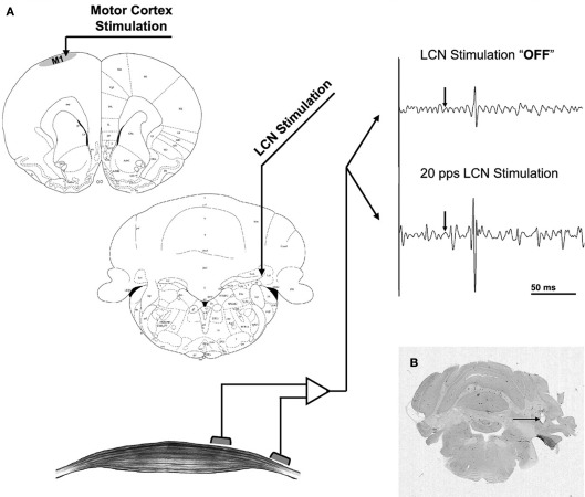
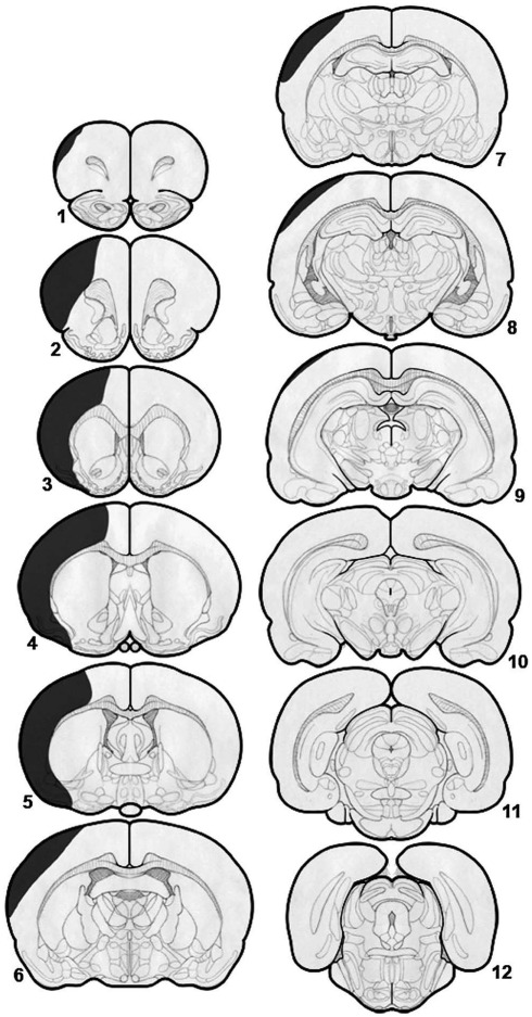
Method: In previous studies, motor evoked potentials were derived from intracortical microstimulation and used to index cortical excitability in rats undergoing continuous, asynchronous deep cerebellar stimulation. In a separate set of experiments, the effect of chronic deep cerebellar stimulation on motor recovery was tested in rats following large ischemic strokes.

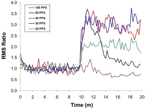
Results: Deep cerebellar stimulation modulated cortical excitability in a frequency-dependent fashion. Beta band stimulation yielded sustained increment in excitability and was associated with enhanced motor recovery compared to sham stimulation.

Conclusion: Chronic deep cerebellar stimulation enhances recovery of motor function following large ischemic strokes in the rat, an effect that may be associated with increased cortical excitability. Given that deep brain stimulation is already a well established method, this new approach to motor recovery may be a viable option for human translation in stroke rehabilitation.

Keywords: deep brain stimulation; diaschisis; electrical stimulation; plasticity; rehabilitation; stroke.


Figures

Figure 1
Figure 1
(A) Stimulation and recording set-up. The intracortical microstimulation electrode was placed in the motor cortex (upper left) while MEPs were recorded from the contralateral hamstrings (bottom left). LCN stimulation was delivered via a macroelectrode (center left) and is not coupled to intracortical microstimulation. The raw EMG tracings (upper right) represent a 200-ms segment, comprised of a 50-ms baseline followed by a 150-ms response window. The arrow represents the time of intracortical stimulation. (B) Coronal cut of the rat’s cerebellum stained for H&E. The artifact corresponding to the tip of the macroelectrode targeted at the LCN can be seen (arrow). With permission from Elsevier Limited 72.
Figure 2
Figure 2
Effects of LCN stimulation on mean MEP amplitude over time, showing frequency dependency of the response. The initial 10 min of data represent the “off” LCN stimulation condition, followed by 10-min of “on.” The effects for each stimulation frequency are shown. All frequency conditions show an initial increase in MEP response magnitude at the start of LCN stimulation, except for 100 Hz. At 50 Hz, the effect is transient, with the response approximating baseline (“off”) levels by the end of the 10-min block. A similar but less dramatic pattern is seen for stimulation at 40 Hz. The enhancement is sustained at both 20 and 30 Hz but more pronounced at 30 Hz. With permission from Elsevier Limited 72.
Figure 3
Figure 3
Diagrammatic representation of a typical stroke associated with coagulation and division of the middle cerebral artery and 30-min occlusion–reperfusion of both common carotid arteries. There is significant damage to the cortex anterior to the bregma but relative sparing of the areas posterior to the bregma. There is almost total sparing of the basal ganglia and thalamus. With permission from Elsevier Limited 85.

References

    1. Abe K., Ukita H., Yorifuji S., Yanagihara T. (1997). Crossed cerebellar diaschisis in chronic Broca’s aphasia. Neuroradiology 39, 624–62610.1007/s002340050480 - DOI - PubMed
    1. Adkins D. L., Campos P., Quach D., Borromeo M., Schallert K., Jones T. A. (2006). Epidural cortical stimulation enhances motor function after sensorimotor cortical infarcts in rats. Exp. Neurol. 200, 356–37010.1016/j.expneurol.2006.02.131 - DOI - PubMed
    1. Adkins D. L., Hsu J. E., Jones T. A. (2008). Motor cortical stimulation promotes synaptic plasticity and behavioral improvements following sensorimotor cortex lesions. Exp. Neurol. 212, 14–2810.1016/j.expneurol.2008.01.031 - DOI - PMC - PubMed
    1. Adkins-Muir D. L., Jones T. A. (2003). Cortical electrical stimulation combined with rehabilitative training: enhanced functional recovery and dendritic plasticity following focal cortical ischemia in rats. Neurol. Res. 25, 780–78810.1179/016164103771953853 - DOI - PubMed
    1. Baker K. B., Schuster D., Cooperrider J., Machado A. G. (2010). Deep brain stimulation of the lateral cerebellar nucleus produces frequency-specific alterations in motor evoked potentials in the rat in vivo. Exp. Neurol. 226, 259–26410.1016/j.expneurol.2010.08.019 - DOI - PMC - PubMed

LinkOut - more resources