. 2012 May 22:3:138.
doi: 10.3389/fphys.2012.00138. eCollection 2012.

Toll-like receptors in liver fibrosis: cellular crosstalk and mechanisms

Affiliations

Toll-like receptors in liver fibrosis: cellular crosstalk and mechanisms

Ling Yang et al. Front Physiol. .

Abstract

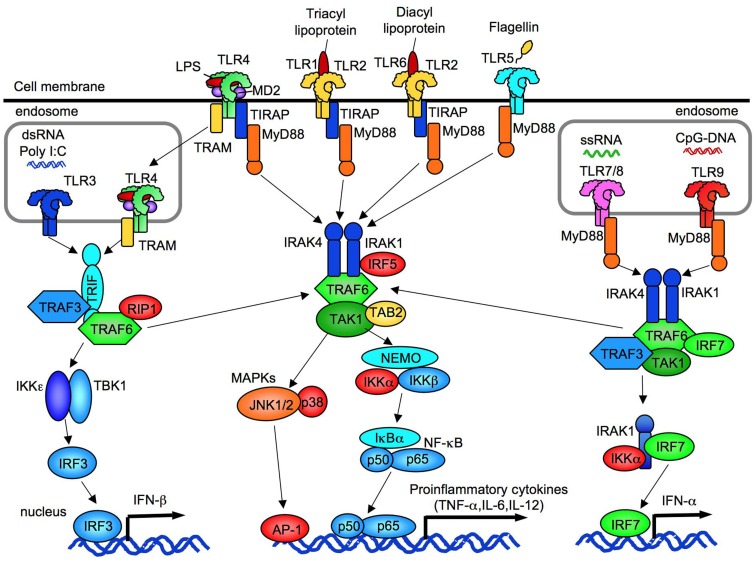
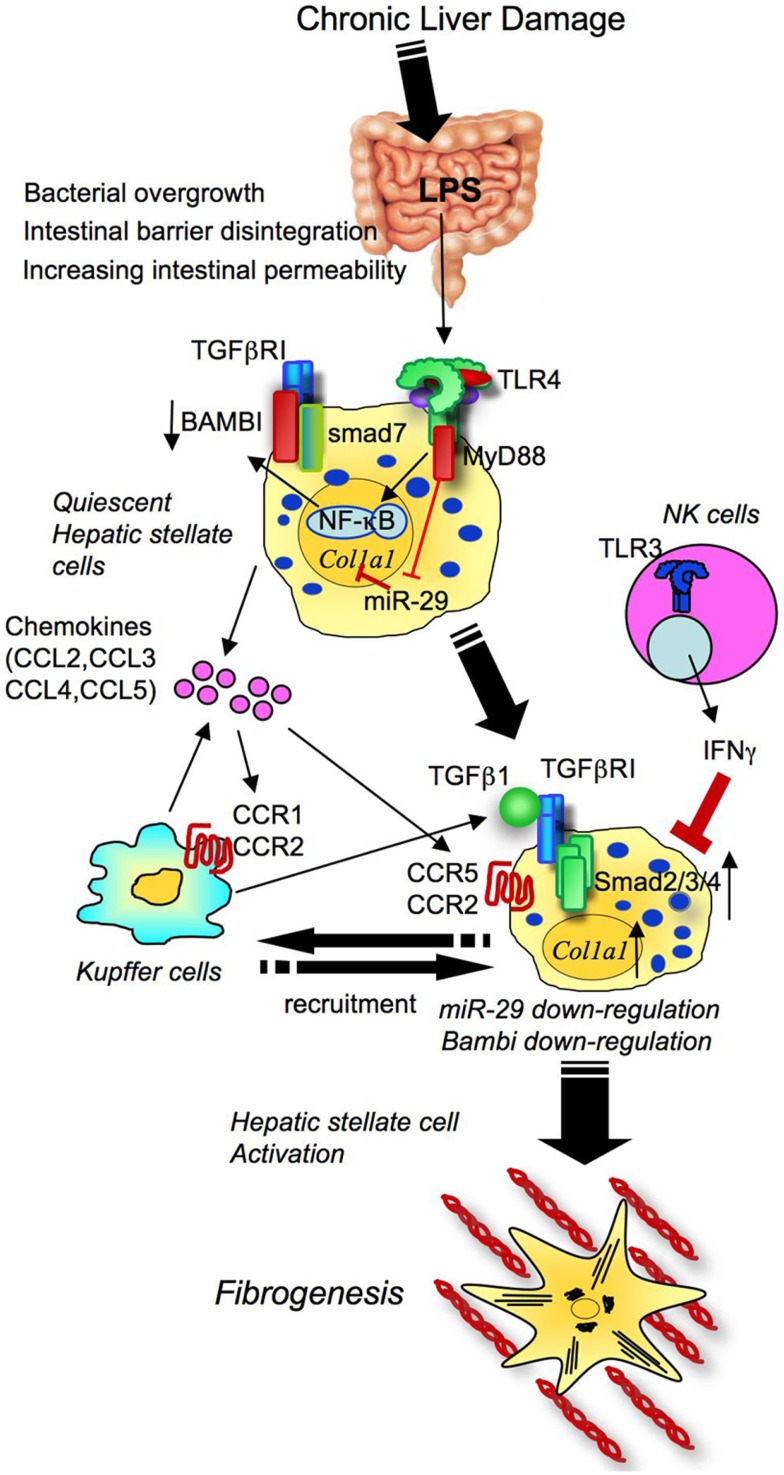
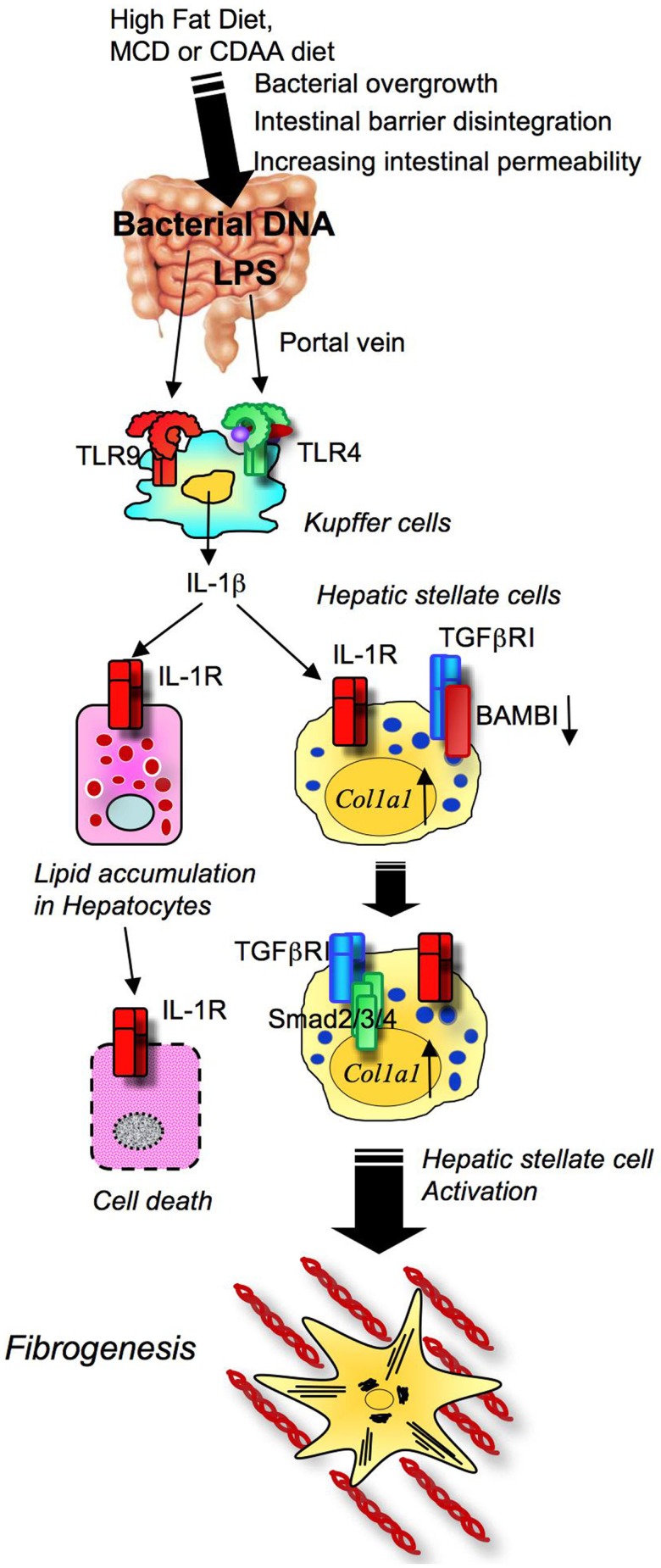
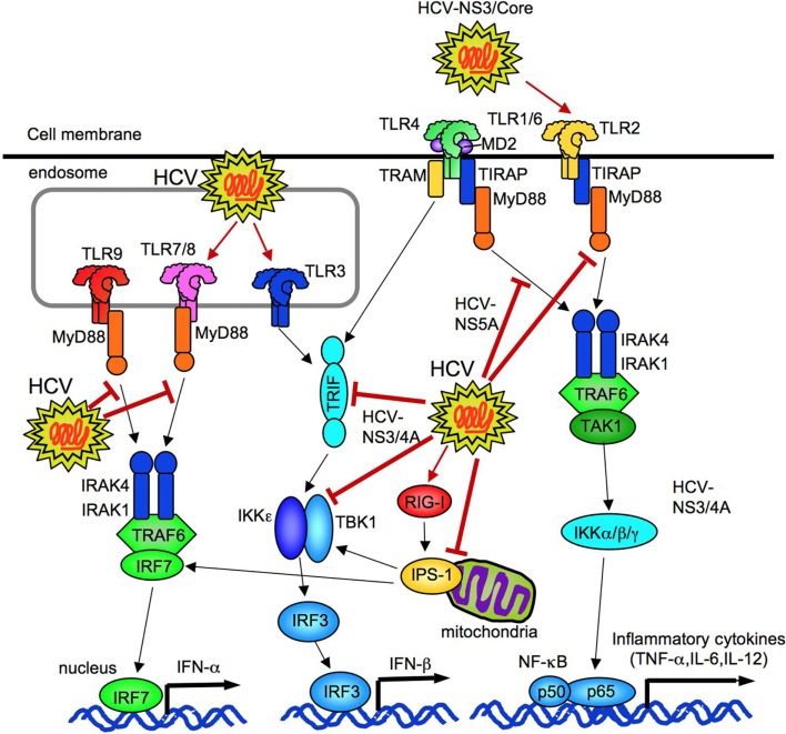
Toll-like receptors (TLRs) are pattern recognition receptors that distinguish conserved microbial products, also known as pathogen-associated molecular patterns (PAMPs), from host molecules. Liver is the first filter organ between the gastrointestinal tracts and the rest of the body through portal circulation. Thus, the liver is a major organ that must deal with PAMPs and microorganisms translocated from the intestine and to respond to the damage associated molecular patterns (DAMPs) released from injured organs. These PAMPs and DAMPs preferentially activate TLR signaling on various cell types in the liver inducing the production of inflammatory and fibrogenic cytokines that initiate and prolong liver inflammation, thereby leading to fibrosis. We summarize recent findings on the role of TLRs, ligands, and intracellular signaling in the pathophysiology of liver fibrosis due to different etiology, as well as to highlight the potential role of TLR signaling in liver fibrosis associated with hepatitis C infection, non-alcoholic and alcoholic steatoheoatitis, primary biliary cirrhosis, and cystic fibrosis.

Keywords: alcoholic liver disease; cystic fibrosis; hepatitis C; non-alcoholic steatohepatitis; primary biliary cirrhosis; toll-like receptors.


Figures

Figure 1
Figure 1
The overview of TLR signaling. TLR1, TLR2, TLR4, TLR5, and TLR6 are expressed on cell membrane. TLR3, TLR7/8, and TLR9 are expressed in endosome. All TLRs, expect for TLR3, activate MyD88-dependent pathway to induce NF-κB and p38/JNK activation. TLR2 and TLR4 signaling require TIRAP and MyD88. TLR3 requires IRIF to activate TBK1/IKKε. Subsequent to TLR4 internalization, TLR4 signaling activates TRAM/TRIF-dependent pathway. TLR3/4-dependent TRIF-dependent signaling induces IRF3 activation and IFN-β production. TLR7/8 and TLR9 induce IFN-α production through IRF7.
Figure 2
Figure 2
Negative regulators of TLR signaling. TLR signaling is suppressed at levels of receptors by ST2, SIGGR/TIR8, RP105, and soluble forms of TLRs. Adaptor molecules are inhibited by SOCS-1, sMyD88, SARM, and PTP1B. SOCS-1 and IRAK-M suppresses IRAKs. A20 blocks TRAF6 and RIP1. IL-6 transcription is negatively regulated by IκBNS, Zc3h12a, and ATF3. ATF3 also blocks IL-12p40 transcription.
Figure 3
Figure 3
Negative regulation of TLR signaling by miRNA. miR-146 negatively regulates the expression of IRAK1, IRAK2, and TRAF6. miR-21 negatively regulates the expression of a IL-12 p35 subunit. miR-155 inhibits the expression of TAB2, RIP1, IKKε, SOCS-1, and TIRAP. miR-223 suppresses the expression of TLR4 and IKKα, and miR-9 inhibits the expression of an NF-κB p50 subunit.
Figure 4
Figure 4
miR-155 and miR-21 tunes TLR4 signaling. TLR4 signaling increases the level of miR-155 that depredates SHIP1, a negative regulator of TLR4 signaling. TLR4 signaling also increases the level of miR-21, which targets PDCD4 mRNA, resulting in increased production of IL-10 as PDCD4 is an inhibitor of IL-10 translation. IL-10 further inhibits miR-155 induction, which in turn, leads to an increase in SHIP1, inhibiting TLR4 signaling.
Figure 5
Figure 5
Toll-like receptor signaling drives liver fibrosis through activation of hepatic stellate cells. In chronic liver damage, intestinal permeability is increased due to systemic inflammation, portal hypertension, intestinal dysbiosis, or tight junction disintegrity, leading to bacterial translocation. Translocated LPS stimulates TLR4 on HSCs. Upon activation of TLR4, HSCs produce chemokines to recruit Kupffer cells through CCR1 and CCR2, and Bambi is downregulated through MyD88 and NF-κB. The fully activated TGF-β signaling eventually induces HSC activation and liver fibrosis. TLR4 also inhibits miR-29 to enhance collagen production. CCR5 is important for HSC recruitment. Generally, the NK cells-TLR3-IFNΓ axis suppresses HSC activation.
Figure 6
Figure 6
Toll-like receptor signaling and NASH development. High fat diet feeding and obesity conditions affect the composition of intestinal microflora and bacterial overgrowth. In NASH, translocated LPS and CpG-DNA stimulates TLR4 and TLR9 on Kupffer cells to produce IL-1β, which induces hepatocyte steatosis and death, and ultimately activates HSC, resulting in fibrosis.
Figure 7
Figure 7
TLR4 signaling in alcoholic steatohepatitis and fibrosis. Excessive alcohol abuse induces changes in the composition of intestinal microbiota and bacterial overgrowth. With tight junction disintegrity, intestinal permeability increases, causing the translocation of gut microflora-derived LPS into the liver through the portal vein. Translocated LPS activates TLR4 on both Kupffer cells and HSCs. TLR4 signaling induces the production of chemokines that promote migration of Kupffer cells and HSCs. The TLR4–TRIF–IRF3 pathway and miR-155 control TLR4 activation in Kupffer cells. These events induce liver inflammation, hepatocyte steatosis, and fibrosis.
Figure 8
Figure 8
HCV regulates innate immune responses. HCV can activate TLR2, TLR3, TLR7, and cytosolic RIG-I. HCV (NS3/4A and NS3) inhibits TRIF and TBK1 to suppress IFN-β production. HCV (NS5A) binds to MyD88 to prevent TLR2, 4, 7, and 9 signaling. HCV (NS3/4A) cleaves of IPS-1 at C508, resulting in prevention of IRF3-mediated IFN-β production.

References

    1. Abe T., Kaname Y., Hamamoto I., Tsuda Y., Wen X., Taguwa S., Moriishi K., Takeuchi O., Kawai T., Kanto T., Hayashi N., Akira S., Matsuura Y. (2007). Hepatitis C virus nonstructural protein 5A modulates the toll-like receptor-MyD88-dependent signaling pathway in macrophage cell lines. J. Virol. 81, 8953–896610.1128/JVI.00649-07 - DOI - PMC - PubMed
    1. Adachi Y., Bradford B. U., Gao W., Bojes H. K., Thurman R. G. (1994). Inactivation of Kupffer cells prevents early alcohol-induced liver injury. Hepatology 20, 453–46010.1002/hep.1840200227 - DOI - PubMed
    1. Adachi Y., Moore L. E., Bradford B. U., Gao W., Thurman R. G. (1995). Antibiotics prevent liver injury in rats following long-term exposure to ethanol. Gastroenterology 108, 218–22410.1016/0016-5085(95)90027-6 - DOI - PubMed
    1. Adawi D., Kasravi F. B., Molin G., Jeppsson B. (1997). Effect of Lactobacillus supplementation with and without arginine on liver damage and bacterial translocation in an acute liver injury model in the rat. Hepatology 25, 642–64710.1002/hep.510250325 - DOI - PubMed
    1. Ambrosini Y. M., Yang G. X., Zhang W., Tsuda M., Shu S., Tsuneyama K., Leung P. S., Ansari A. A., Coppel R. L., Gershwin M. E. (2011). The multi-hit hypothesis of primary biliary cirrhosis: polyinosinic-polycytidylic acid (poly I:C) and murine autoimmune cholangitis. Clin. Exp. Immunol. 166, 110–12010.1111/j.1365-2249.2011.04453.x - DOI - PMC - PubMed