

MRI tracking of FePro labeled fresh and cryopreserved long term in vitro expanded human cord blood AC133+ endothelial progenitor cells in rat glioma
- PMID: 22662174
- PMCID: PMC3360770
- DOI: 10.1371/journal.pone.0037577
MRI tracking of FePro labeled fresh and cryopreserved long term in vitro expanded human cord blood AC133+ endothelial progenitor cells in rat glioma
Abstract
Background: Endothelial progenitors cells (EPCs) are important for the development of cell therapies for various diseases. However, the major obstacles in developing such therapies are low quantities of EPCs that can be generated from the patient and the lack of adequate non-invasive imaging approach for in vivo monitoring of transplanted cells. The objective of this project was to determine the ability of cord blood (CB) AC133+ EPCs to differentiate, in vitro and in vivo, toward mature endothelial cells (ECs) after long term in vitro expansion and cryopreservation and to use magnetic resonance imaging (MRI) to assess the in vivo migratory potential of ex vivo expanded and cryopreserved CB AC133+ EPCs in an orthotopic glioma rat model.
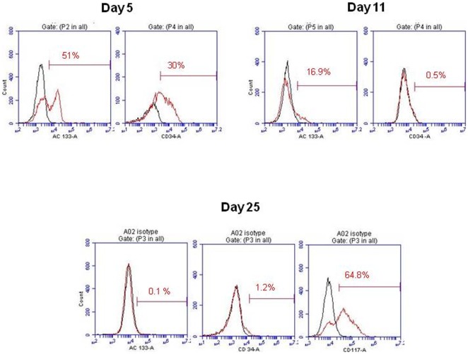
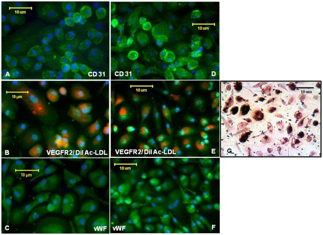
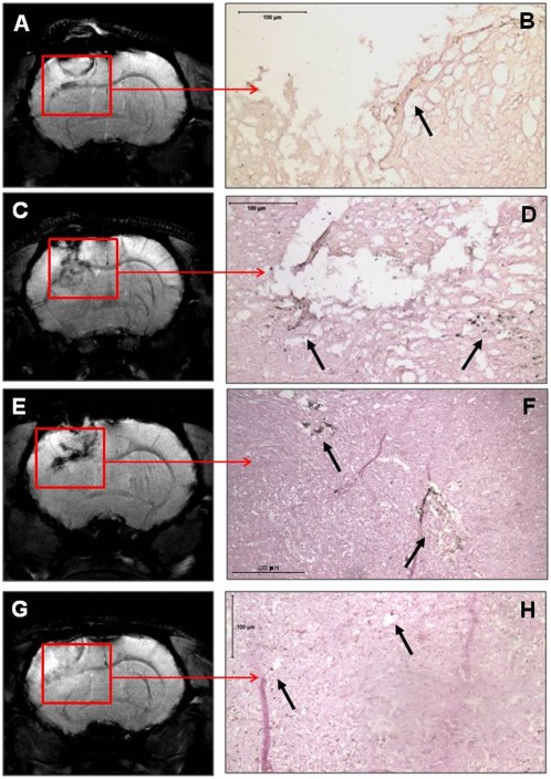
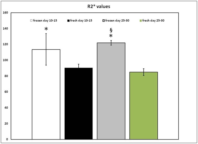
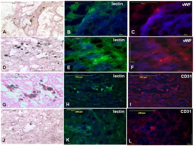
Materials, methods and results: The primary CB AC133+ EPC culture contained mainly EPCs and long term in vitro conditions facilitated the maintenance of these cells in a state of commitment toward endothelial lineage. At days 15-20 and 25-30 of the primary culture, the cells were labeled with FePro and cryopreserved for a few weeks. Cryopreserved cells were thawed and in vitro differentiated or i.v. administered to glioma bearing rats. Different groups of rats also received long-term cultured, magnetically labeled fresh EPCs and both groups of animals underwent MRI 7 days after i.v. administration of EPCs. Fluorescent microscopy showed that in vitro differentiation of EPCs was not affected by FePro labeling and cryopreservation. MRI analysis demonstrated that in vivo accumulation of previously cryopreserved transplanted cells resulted in significantly higher R2 and R2* values indicating a higher rate of migration and incorporation into tumor neovascularization of previously cryopreserved CB AC133+ EPCs to glioma sites, compared to non-cryopreserved cells.
Conclusion: Magnetically labeled CB EPCs can be in vitro expanded and cryopreserved for future use as MRI probes for monitoring the migration and incorporation to the sites of neovascularization.
Conflict of interest statement
Figures
Similar articles
-
Human cord blood-derived AC133+ progenitor cells preserve endothelial progenitor characteristics after long term in vitro expansion.PLoS One. 2010 Feb 11;5(2):e9173. doi: 10.1371/journal.pone.0009173. PLoS One. 2010. PMID: 20161785 Free PMC article.
-
Magnetic resonance imaging and confocal microscopy studies of magnetically labeled endothelial progenitor cells trafficking to sites of tumor angiogenesis.Stem Cells. 2006 Mar;24(3):671-8. doi: 10.1634/stemcells.2005-0017. Epub 2005 Sep 22. Stem Cells. 2006. PMID: 16179427
-
Endothelial progenitor cells (EPCs) as gene carrier system for rat model of human glioma.PLoS One. 2012;7(1):e30310. doi: 10.1371/journal.pone.0030310. Epub 2012 Jan 20. PLoS One. 2012. PMID: 22276177 Free PMC article.
-
Endothelial progenitor cells in angiogenesis.Sheng Li Xue Bao. 2005 Feb 25;57(1):1-6. Sheng Li Xue Bao. 2005. PMID: 15719128 Review.
-
Endothelial progenitor cells: mobilization, differentiation, and homing.Arterioscler Thromb Vasc Biol. 2003 Jul 1;23(7):1185-9. doi: 10.1161/01.ATV.0000073832.49290.B5. Epub 2003 Apr 24. Arterioscler Thromb Vasc Biol. 2003. PMID: 12714439 Review.
Cited by
-
Differential biodistribution of intravenously administered endothelial progenitor and cytotoxic T-cells in rat bearing orthotopic human glioma.BMC Med Imaging. 2013 Jun 10;13:17. doi: 10.1186/1471-2342-13-17. BMC Med Imaging. 2013. PMID: 23758888 Free PMC article.
-
Application of Umbilical Cord Blood Derived Stem Cells in Diseases of the Nervous System.J Stem Cell Res Ther. 2014;4:1000202. doi: 10.4172/2157-7633.1000202. J Stem Cell Res Ther. 2014. PMID: 25599002 Free PMC article.
-
Changes in the tumor microenvironment and outcome for TME-targeting therapy in glioblastoma: A pilot study.PLoS One. 2021 Feb 5;16(2):e0246646. doi: 10.1371/journal.pone.0246646. eCollection 2021. PLoS One. 2021. PMID: 33544755 Free PMC article.
-
Antiangiogenic Targets for Glioblastoma Therapy from a Pre-Clinical Approach, Using Nanoformulations.Int J Mol Sci. 2020 Jun 24;21(12):4490. doi: 10.3390/ijms21124490. Int J Mol Sci. 2020. PMID: 32599834 Free PMC article. Review.
-
Intravenous administration of human umbilical cord blood-derived AC133+ endothelial progenitor cells in rat stroke model reduces infarct volume: magnetic resonance imaging and histological findings.Stem Cells Transl Med. 2013 Sep;2(9):703-14. doi: 10.5966/sctm.2013-0066. Epub 2013 Aug 9. Stem Cells Transl Med. 2013. PMID: 23934909 Free PMC article.
References
-
- Scott SM, Barth MG, Gaddy LR, Ahl ET The role of circulating cells in the healing of vascular prostheses. J Vasc Surg. 1994;19:585–593. - PubMed
-
- Solovey A, Lin Y, Browne P, Choong S, Wayner E, et al. Circulating activated endothelial cells in sickle cell anemia. N Engl J Med. 1997;337:1584–1590. - PubMed
-
- Sowemimo-Coker SO, Meiselman HJ, Francis RB Increased circulating endothelial cells in sickle cell crisis. Am J Hematol. 1989;31:263–265. - PubMed
-
- Hladovec J. Circulating endothelial cells as a sign of vessel wall lesions. Physiol Bohemoslov. 1978;27:140–144. - PubMed
-
- Hladovec J, Prerovsky I, Stanek V, Fabian J. Circulating endothelial cells in acute myocardial infarction and angina pectoris. Klin Wochenschr. 1978;56:1033–1036. - PubMed
Publication types
MeSH terms
Substances
Grants and funding
LinkOut - more resources
Full Text Sources
Medical
Research Materials
