. 2012;7(5):e37628.
doi: 10.1371/journal.pone.0037628. Epub 2012 May 25.

Functional analysis of a breast cancer-associated mutation in the intracellular domain of the metalloprotease ADAM12

Affiliations

Functional analysis of a breast cancer-associated mutation in the intracellular domain of the metalloprotease ADAM12

Dorte Stautz et al. PLoS One. 2012.

Abstract

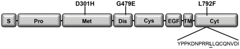
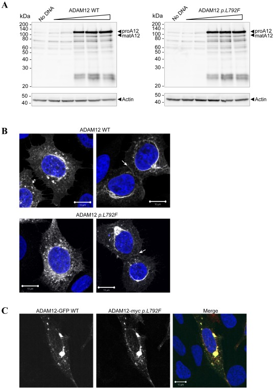
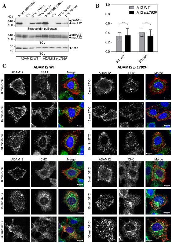
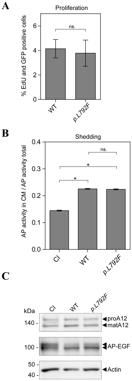
A recently identified breast cancer-associated mutation in the metalloprotease ADAM12 alters a potential dileucine trafficking signal, which could affect protein processing and cellular localization. ADAM12 belongs to the group of A Disintegrin And Metalloproteases (ADAMs), which are typically membrane-associated proteins involved in ectodomain shedding, cell-adhesion, and signaling. ADAM12 as well as several members of the ADAM family are over-expressed in various cancers, correlating with disease stage. Three breast cancer-associated somatic mutations were previously identified in ADAM12, and two of these, one in the metalloprotease domain and another in the disintegrin domain, were investigated and found to result in protein misfolding, retention in the secretory pathway, and failure of zymogen maturation. The third mutation, p.L792F in the ADAM12 cytoplasmic tail, was not investigated, but is potentially significant given its location within a di-leucine motif, which is recognized as a potential cellular trafficking signal. The present study was motivated both by the potential relevance of this documented mutation to cancer, as well as for determining the role of the di-leucine motif in ADAM12 trafficking. Expression of ADAM12 p.L792F in mammalian cells demonstrated quantitatively similar expression levels and zymogen maturation as wild-type (WT) ADAM12, as well as comparable cellular localizations. A cell surface biotinylation assay demonstrated that cell surface levels of ADAM12 WT and ADAM12 p.L792F were similar and that internalization of the mutant occurred at the same rate and extent as for ADAM12 WT. Moreover, functional analysis revealed no differences in cell proliferation or ectodomain shedding of epidermal growth factor (EGF), a known ADAM12 substrate between WT and mutant ADAM12. These data suggest that the ADAM12 p.L792F mutation is unlikely to be a driver (cancer causing)-mutation in breast cancer.


Conflict of interest statement

Competing Interests: The authors have declared that no competing interests exist.

Figures

Figure 1
Figure 1. Schematic illustration of ADAM12 indicating the published breast cancer-associated mutations and the di-leucine motif in the cytoplasmic tail.
Schematic illustration of the domain organization of ADAM12. S: Signal peptide; Pro: Prodomain; Met: Metalloprotease domain; Dis: Disintegrin domain; Cys: Cysteine-rich domain; EGF: EGF-like domain; TM: Transmembrane domain; Cyt: Cytoplasmic tail. Positions of the three breast cancer-associated mutations are indicated and the amino acid sequence containing the di-leucine motif is shown.
Figure 2
Figure 2. Expression and processing of ADAM12 wild-type and p.L792F mutant.
(A) ADAM12 p.L792F is expressed and processed similarly to WT ADAM12. 293VnR cells were transiently transfected with increasing amounts of either ADAM12 WT or ADAM12 p.L792F. After 48 h cells were lysed, total protein amounts were determined and proteins analyzed by SDS-PAGE and subsequent western blot with an anti-ADAM12 antibody. The ADAM12 proform (proA12) is ∼120 kDa and the mature form (matA12) is ∼95 kDa (arrowheads). The membranes were re-probed with anti-actin antibody to confirm equal loading. (B) Similar immunofluorescent staining of ADAM12 WT and p.L792F mutant. 293VnR cells were transiently transfected with ADAM12 WT or ADAM12 p.L792F and after two days of incubation, seeded onto FBS coated coverslips. Cells were fixed in 4% PFA, permeabilized in Triton X-100, labeled with ADAM12 monoclonal antibody 6E6 for 1 hour, and followed by staining with secondary antibody and DAPI. Two representative images acquired using confocal laser-scanning microscopy are shown for each protein. White arrows indicate staining at cell-cell junctions. Scale bar = 10 µm. (C) 293VnR cells were transiently transfected with ADAM12-GFP WT or ADAM12-myc p.L792F and processed as described in B, except that cells were labeled with anti-myc instead of anti-ADAM12 primary antibody. Scale bar = 10 µm.
Figure 3
Figure 3. ADAM12 p.L792F localizes to the cell surface and is internalized similarly to wild-type ADAM12.
(A) Biotinylation of cell surface ADAM12 WT or ADAM12 p.L792F. 293VnR cells, transiently transfected with ADAM12-GFP or ADAM12-GFP p.L792F were labeled with NHS-SS-Biotin for 30 min at 4°C, with subsequent incubation at 4°C (controls) or at 37°C for indicated time points. Then, the cells were treated with reducing reagent (glutathione) to remove non-internalized biotin from the cell surface (except for total biotinylation control lane 1). The control samples kept at 4°C were included to ensure proper removal of cell surface biotin. Biotinylated proteins were precipitated with streptavidin-conjugated agarose beads and subjected to western blotting with the appropriate antibodies. The ADAM12-GFP proform (proA12) is ∼150 kDa and the mature form (matA12) is ∼130 kDa. Actin was used as a loading control. TCL: Total cell lysate. (B) Quantitative analysis of ADAM12 internalization where internalized ADAM12-GFP at 30 or 60 min at 37°C were calculated as fold-change compared to total biotinylated ADAM12-GFP for each WT or mutant sample. The data are shown as +/− standard error of the mean (SEM) (n = 5 independent experiments); “ns.” indicates no statistical difference (p>0.05, two-tailed unpaired t-test). (C) ADAM12 WT or ADAM12 p.L792F internalization and co-localization with the early endosomal marker (EEA1) and clathrin heavy chain (CHC). The images show immunofluorescent staining and confocal laser-scanning microscopy of 293VnR cells transiently transfected with ADAM12 WT or the p.L792F mutant and seeded onto FBS-coated coverslips. Cells were labeled with ADAM12 monoclonal antibody 6E6 for 1 hour at 4°C, washed, and incubated in culture medium for 15 min or 30 min, followed by fixation and permeabilization. Cells were then stained with an EEA1 antibody (top three panels) or CHC antibody (bottom three panels) for 1 h at RT and subsequently with appropriate secondary antibodies and DAPI (n = 2). Merged images show partial co-localization of ADAM12 and EEA1 or CHC. Scale bar = 10 µm.
Figure 4
Figure 4. ADAM12 p.L792F does not affect cell proliferation or EGF shedding.
(A) Comparable proliferation of ADAM12 WT and ADAM12 p.L792F expressing cells. 293VnR cells transiently transfected with ADAM12-GFP or ADAM12-GFP p.L792F were plated on FBS-coated coverslips in triplicates. Proliferating cells were stained with Edu. Four images were taken from each triplicate and EdU- and GFP-positive cells were calculated as percentage of total cells (DAPI staining). The data are shown as +/− standard error of the mean (SEM) (n = 2 independent experiments performed in triplicates); “ns.” indicates no statistical difference (p>0.05, two-tailed unpaired t-test). (B) ADAM12 p.L792F does not affect ADAM12-mediated proEGF shedding. 293VnR cells were transiently transfected with alkaline phosphatase (AP)-tagged proEGF together with ADAM12-GFP WT, CI ADAM12-GFP, or ADAM12 p.L792F (p.L792F). EGF-shedding was calculated from the ratio of AP activity in the cell media to total AP activity in the media and corresponding cell lysate, corrected for background (sample without AP substrate) (n = 2 independent experiments performed in triplicate). * indicates a statistically significant difference (p<0.0001, two-tailed unpaired t-test) and “ns.” indicates no statistical difference (p>0.05). (C) Representative western blot of protein expression control from experiment in (B). Transfected 293VnR cells were lysed in RIPA buffer and analyzed by western blot with the appropriate antibodies. Note the reduction in the upper band of AP-EGF, indicating shedding of proEGF-AP from the cell surface. Actin was used as a loading control.

Similar articles

Cited by

References

    1. Edwards DR, Handsley MM, Pennington CJ. The ADAM metalloproteinases. Mol Aspects Med. 2008;29:258–289. - PMC - PubMed
    1. Blobel CP, Carpenter G, Freeman M. The role of protease activity in ErbB biology. Exp Cell Res. 2009;315:671–682. - PMC - PubMed
    1. Murphy G. The ADAMs: signalling scissors in the tumour microenvironment. Nat Rev Cancer. 2008;8:929–941. - PubMed
    1. Murphy G. Fell-Muir Lecture: Metalloproteinases: from demolition squad to master regulators. Int J Exp Pathol. 2010;91:303–313. - PMC - PubMed
    1. Iba K, Albrechtsen R, Gilpin BJ, Loechel F, Wewer UM. Cysteine-rich domain of human ADAM 12 (meltrin alpha) supports tumor cell adhesion. Am J Pathol. 1999;154:1489–1501. - PMC - PubMed

Publication types