. 2012;7(5):e37746.
doi: 10.1371/journal.pone.0037746. Epub 2012 May 25.

Lipoxin A4 stimulates calcium-activated chloride currents and increases airway surface liquid height in normal and cystic fibrosis airway epithelia

Affiliations

Lipoxin A4 stimulates calcium-activated chloride currents and increases airway surface liquid height in normal and cystic fibrosis airway epithelia

Valia Verrière et al. PLoS One. 2012.

Abstract

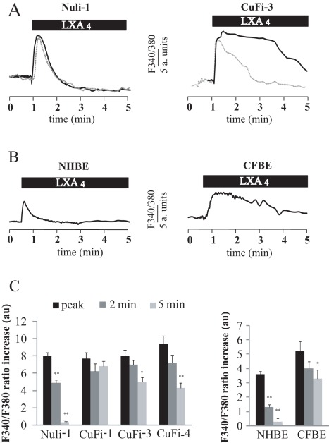
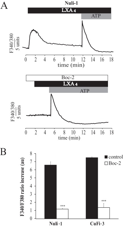
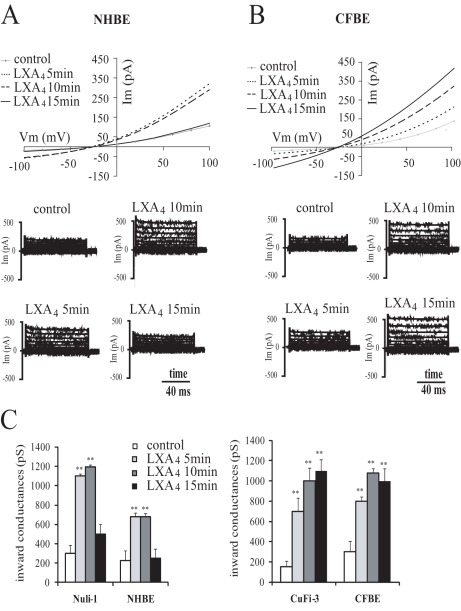
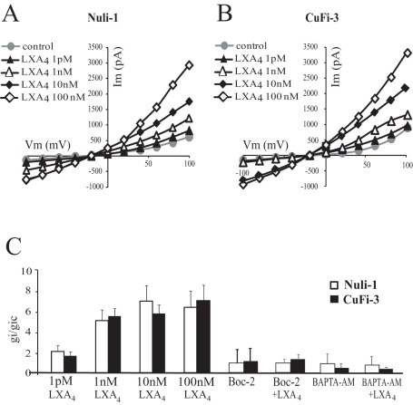
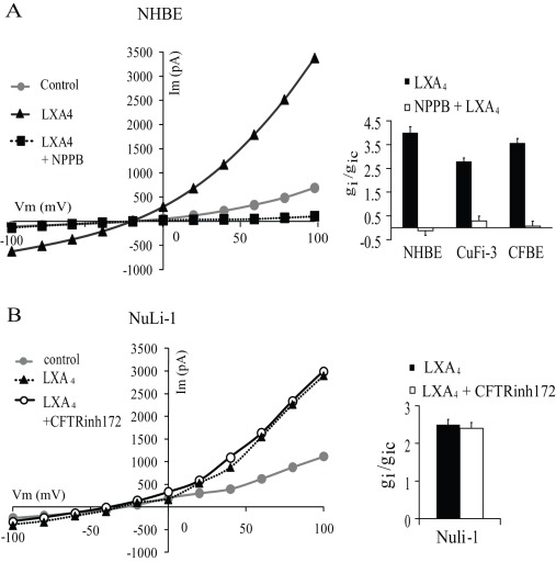
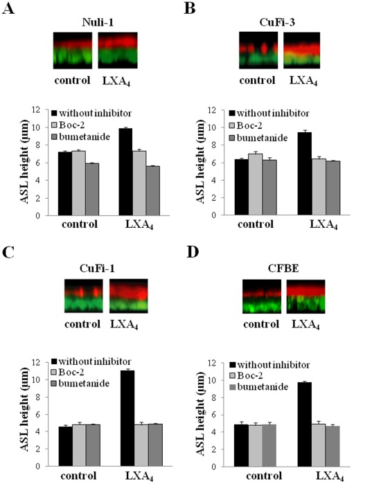
Cystic Fibrosis (CF) is a genetic disease characterised by a deficit in epithelial Cl(-) secretion which in the lung leads to airway dehydration and a reduced Airway Surface Liquid (ASL) height. The endogenous lipoxin LXA(4) is a member of the newly identified eicosanoids playing a key role in ending the inflammatory process. Levels of LXA(4) are reported to be decreased in the airways of patients with CF. We have previously shown that in normal human bronchial epithelial cells, LXA(4) produced a rapid and transient increase in intracellular Ca(2+). We have investigated, the effect of LXA(4) on Cl(-) secretion and the functional consequences on ASL generation in bronchial epithelial cells obtained from CF and non-CF patient biopsies and in bronchial epithelial cell lines. We found that LXA(4) stimulated a rapid intracellular Ca(2+) increase in all of the different CF bronchial epithelial cells tested. In non-CF and CF bronchial epithelia, LXA(4) stimulated whole-cell Cl(-) currents which were inhibited by NPPB (calcium-activated Cl(-) channel inhibitor), BAPTA-AM (chelator of intracellular Ca(2+)) but not by CFTRinh-172 (CFTR inhibitor). We found, using confocal imaging, that LXA(4) increased the ASL height in non-CF and in CF airway bronchial epithelia. The LXA(4) effect on ASL height was sensitive to bumetanide, an inhibitor of transepithelial Cl(-) secretion. The LXA(4) stimulation of intracellular Ca(2+), whole-cell Cl(-) currents, conductances and ASL height were inhibited by Boc-2, a specific antagonist of the ALX/FPR2 receptor. Our results provide, for the first time, evidence for a novel role of LXA(4) in the stimulation of intracellular Ca(2+) signalling leading to Ca(2+)-activated Cl(-) secretion and enhanced ASL height in non-CF and CF bronchial epithelia.


Conflict of interest statement

Competing Interests: The authors have declared that no competing interests exist.

Figures

Figure 1
Figure 1. LXA4 effect on intracellular Ca2+ activity in non-CF and CF bronchial epithelial cells.
(A) Typical effect of LXA4 (100 nM) on the cytosolic Ca2+ (ratio F340/F380) measured in Nuli-1 and CuFi-3 cell lines in control (plain line) and in external Ca2+ -free conditions (dotted line). (B) Typical effect of LXA4 (100 nM) on the cytosolic Ca2+ in normal and CF primary cultures of bronchial epithelial cells (NHBE and CFBE). (C) Mean values of the maximum increase in Ca2+ (peak) and measured 2 and 5 minutes after the peak, in Nuli-1 (n = 6), CuFi-1 (n = 6), CuFi-3 (n = 6), CuFi-4 (n = 4) cell lines and in NHBE (n = 4) and CFBE (n = 4) bronchial epithelial cells in primary culture (* p<0.05, ** p<0.01).
Figure 2
Figure 2. Effect of Boc-2 on the intracellular Ca2+ signal induced by LXA4.
(A) Representative effect of LXA4 (100 nM) and ATP (100 µM) on cytosolic Ca2+ (ratio F340/F380) in NuLi-1 cells in control conditions (upper panel) and after 24 hours of pre-treatment with Boc-2 (10 µM) a specific inhibitor of ALX/FPR2 (lower panel). (B) Mean values corresponding to the effect of Boc-2 on the Ca2+ response to LXA4 in Nuli-1 (n = 5) and in CuFi-3 (n = 4) cell lines (*** p<0.001).
Figure 3
Figure 3. Time dependency of the effect of LXA4 on whole-cell currents in normal and CF bronchial epithelial cells in primary culture.
Typical I-V relationships and corresponding current records obtained before and after 5, 10 and 15 min exposure to LXA4 (100 nM) in NHBE (A) and CFBE (B) isolated bronchial epithelial cells in primary culture. (C) Mean values corresponding to the time dependency of the LXA4 effect onwhole cell inward conductances in Nuli-1 (n = 8) and in CuFi-3 (n = 4) cell lines and NHBE (n = 8) and CFBE (n = 6) primary cultures (**p<0.01).
Figure 4
Figure 4. Dose dependency of the effect of LXA4 on whole-cell currents of normal (Nuli-1) and CF (CuFi-3) bronchial epithelial cell lines.
Typical I-V relationships obtained before and after 10 min exposure to 1 pM (n = 6 Nuli-1, n = 6 CuFi-3), 1 nM (n = 4 Nuli-1, n = 4 CuFi-3), 10 nM (n = 3 Nuli-1, n = 3 CuFi-3) and 100 nM (n = 6 Nuli-1, n = 6 CuFi-3) LXA4 in Nuli-1 (A) and CuFi-3 (B) cell lines. (C) Mean inward conductance changes normalized to control values (gi/gic obtained without LXA4) as a function of LXA4 concentration in Nuli-1 (open bars) and CuFi-3 (black bars) cells in control conditions and obtained upon exposure to Boc-2 (10 µM) alone (10 min, 100 nM, n = 4 Nuli-1, n = 4 CuFi-3) or with Boc-2 (10 µM) and LXA4 (10 min, 100 nM, n = 6 Nuli-1, n = 6 CuFi-3) and after BAPTA-AM pre-treatment alone (n = 6 Nuli-1, n = 6 CuFi-3) or with BAPTA-AM and LXA4 (10 min, 100 nM, n = 4 Nuli-1, n = 6 CuFi-3).
Figure 5
Figure 5. Effects of Cl channel inhibitors on whole-cell currents stimulated by LXA4.
(A) Representative I-V relationship of the effect of NPPB (1 µM) after the stimulation of primary cultures of bronchial epithelial cells with LXA4 (100 nM). Corresponding histogram of the inhibitory effect of NPPB on the inward conductance obtained in non-CF primary NHBE cells (n = 4), CuFi-3 cells (n = 3) and CF primary CFBE cells (n = 3) after stimulation with LXA4 (100 nM, 10 min). (B). Typical I-V relationships of the effect of LXA4 (100 nM) obtained in NuLi-1 cells after treatment with the CFTR channel inhibition CFTR-inh172 (5 mM) and histogram showing the absence of inhibitory effect in Nuli-1 cells (n = 3).
Figure 6
Figure 6. LXA4 (100 nM) effects on airway surface liquid height in NuLi-1 (A), CuFi-3 (B) and CuFi-1 epithelial cell line (C) and CFBE primary cultures (D).
Epithelial cells were stained with calcein green, and the ASL labelled with dextran-conjugated Texas red™ fluorochrome. For each cell preparation, typical z-plane confocal sections showing ASL responses to LXA4 (top) and mean ASL height changes (bottom) in control conditions or following LXA4 (100 µM) exposure in NuLi-1,CuFi-3, CuFi-1 and CFBE epithelia treated or not with Boc-2 (10 µM) and with bumetanide (10 µM).

References

    1. Boat TF, Cheng PW. Epithelial cell dysfunction in cystic fibrosis: implications for airways disease. Acta Paediatr Scand Suppl 363: 25–29; discussion. 1989;29–30 - PubMed
    1. Mickle JE, Macek M, Fulmer-Smentek SB, Egan MM, Schwiebert E, et al. A mutation in the cystic fibrosis transmembrane conductance regulator gene associated with elevated sweat chloride concentrations in the absence of cystic fibrosis. Hum Mol Genet. 1998;7:729–735. - PubMed
    1. Davis PB, Drumm M, Konstan MW. Cystic fibrosis. Am J Respir Crit Care Med. 1996;154:1229–1256. - PubMed
    1. Karp CL, Flick LM, Park KW, Softic S, Greer TM, et al. Defective lipoxin-mediated anti-inflammatory activity in the cystic fibrosis airway. Nat Immunol. 2004;5:388–392. - PubMed
    1. Serhan CN, Takano T, Gronert K, Chiang N, Clish CB. Lipoxin and aspirin-triggered 15-epi-lipoxin cellular interactions anti-inflammatory lipid mediators. Clin Chem Lab Med. 1999;37:299–309. - PubMed

Publication types

MeSH terms