. 2012;7(5):e37819.
doi: 10.1371/journal.pone.0037819. Epub 2012 May 25.

Regulation of RKIP function by Helicobacter pylori in gastric cancer

Affiliations

Regulation of RKIP function by Helicobacter pylori in gastric cancer

Erika L Moen et al. PLoS One. 2012.

Abstract

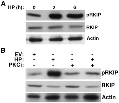
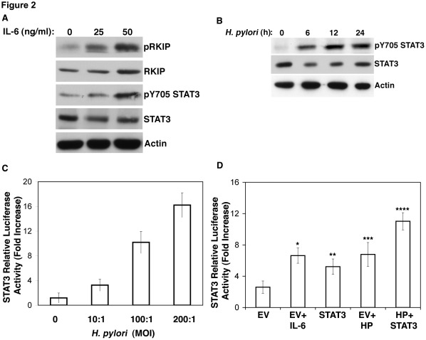
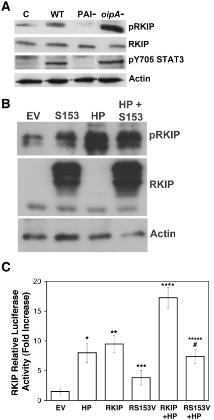
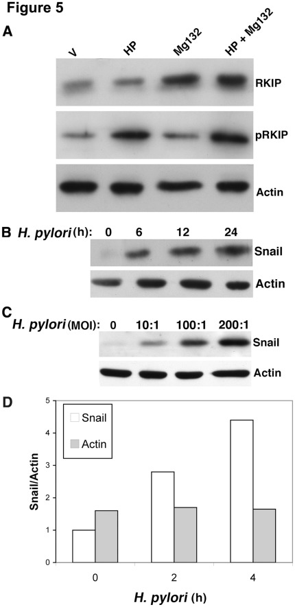
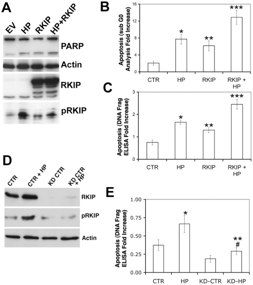
Helicobacter pylori (H. pylori) is a gram-negative, spiral-shaped bacterium that infects more than half of the world's population and is a major cause of gastric adenocarcinoma. The mechanisms that link H. pylori infection to gastric carcinogenesis are not well understood. In the present study, we report that the Raf-kinase inhibitor protein (RKIP) has a role in the induction of apoptosis by H. pylori in gastric epithelial cells. Western blot and luciferase transcription reporter assays demonstrate that the pathogenicity island of H. pylori rapidly phosphorylates RKIP, which then localizes to the nucleus where it activates its own transcription and induces apoptosis. Forced overexpression of RKIP enhances apoptosis in H. pylori-infected cells, whereas RKIP RNA inhibition suppresses the induction of apoptosis by H. pylori infection. While inducing the phosphorylation of RKIP, H. pylori simultaneously targets non-phosphorylated RKIP for proteasome-mediated degradation. The increase in RKIP transcription and phosphorylation is abrogated by mutating RKIP serine 153 to valine, demonstrating that regulation of RKIP activity by H. pylori is dependent upon RKIP's S153 residue. In addition, H. pylori infection increases the expression of Snail, a transcriptional repressor of RKIP. Our results suggest that H. pylori utilizes a tumor suppressor protein, RKIP, to promote apoptosis in gastric cancer cells.


Conflict of interest statement

Competing Interests: The authors have declared that no competing interests exist.

Figures

Figure 1
Figure 1. H. pylori infection results in RKIP phosphorylation.
(A) Western blot analysis of AGS cells co-cultured with H. pylori (HP) at MOI of 100∶1 for 2 and 6 h and examined for pRKIP, RKIP, and actin expression. Densitometry was performed on three independent experiments and band intensities normalized in comparison to Actin for each time point. Our results indicated a 3.126 fold increase (average intensity 0.44 vs 1.376) of pRKIP after 2 h and 1.384 fold increase (average intensity 0.6774 vs 0.938) of RKIP after 2 h of H. pylori infection. (B) AGS cells co-cultured for 6 h in the presence or absence of the PKC inhibitor bisindolylmaleimide (Bis), were examined for the expression of pRKIP, RKIP and actin.All treatments were performed in 1% DMSO as a vehicle control (Bis).
Figure 2
Figure 2. IL-6 promotes RKIP and STAT 3 transcription and phosphorylation; H. pylori infection induces STAT3 transcription and phosphorylation.
(A) Western blot analysis of AGS cells treated with the indicated concentrations of IL-6 and for 6 hours. Densitometry was performed and for pRKIP expression, our results indicate a 1.8 fold incerase of pRKIP (average intensity 0.59 vs 1.051) in cells treated with 25 ng/ml IL-6 and a 1.35 fold increase (average intensity 0.59 vs 0.772) in cells treated with 50 ng/ml IL-6. For RKIP expression, our results indicate a 0.8 fold incerase of pRKIP (average intensity 0.69 vs 0.563) in cells treated with 25 ng/ml IL-6 and a 1.05 fold increase (average intensity 0.69 vs 0.745) in cells treated with 50 ng/ml IL-6 when normalized to actin at each time point. (B) Western blot analysis of AGS co-cultured with H. pylori and examined for pY705 STAT3 for the indicated times; (C) STAT3 luciferase reporter transcriptional assay of AGS cells co-cultured with H. pylori at the indicated MOI; (D) STAT3 luciferase reporter assay of AGS cells transiently transfected with STAT3 for 24 h and co-cultured with H. pylori and/or treated with IL-6 for 6 h. A paired t-test was performed to analyze the increase or decrease in STAT3 transcription of experimental samples when compared to empty vector (EV): *IL-6, p<0.0003; **STAT3, p<0.009; *** H. pylori p<0.0005; **** STAT3 and H. pylori p<0.0000023.
Figure 3
Figure 3. H. pylori infection results in increased RKIP transcription and nuclear localization.
(A) RKIP transcription reporter assay of AGS cells transiently transfected with RKIP luciferase construct and HA-RKIP for 24 h, then co-cultured with H. pylori for 12 h in the presence or absence of the PKC inhibitor. In comparison to empty vector controls, relative transcriptional activity was significantly increased for * H. pylori, p<0.002; and ** H. pylori+RKIP, P<0.0003. Comparing the loss of relative luciferase activity of H. pylori and RKIP when compared to H. pylori, RKIP and Bis, p<0.0004. Data represents the mean +/− standard deviation (sd) of the fold increase relative to empty vector controls in 2 independent experiments performed in duplicate. (B) Western blot analysis of nuclear and cytosolic fractions of AGS cells co-cultured with H. pylori for 4 h for the expression of pRKIP and total RKIP. Actin and laminA provide verification of successful cytoplasmic and nuclear fraction separation.
Figure 4
Figure 4. The pathogenicity island of H. pylori is responsible for RKIP phosphorylation.
Western blot analysis of (A) AGS cells co-cultured with H. pylori strains for 6 h and examined for the indicated proteins. C = control (uninfected), WT = AGS cells infected with wild type H. pylori for 6 h, PAI- and oipA- represent isogenic mutants lacking these genes. (B) AGS cells transiently transfected for 24 h with RKIP S153 cDNA or 24 h and co-cultured with H. pylori for 6 h. (C) RKIP luciferase reporter assay of AGS cells transiently transfected with S153V RKIP in the presence or absence of H. pylori infection. In comparison to empty vector controls, the relative activity of RKIP transcription was increased by: *H. pylori, p<0.002; **RKIP, p<0.002; ***S153V, p<0.03, ****H. pylori and RKIP, p<0.0005; *****H. pylori and S153V, p<0.003. **, RKIP transcriptional activity was significantly decreased by the S153V compared with the wild type RKIP construct in response to H. pylori, #, p<0.0003. The data represents the mean +/− sd of 2 independent experiments performed in duplicate.
Figure 5
Figure 5. H. pylori targets RKIP for proteasomal degradation and results in the induction of Snail.
AGS cells were (A) treated with MG132 and examined for the indicated proteins via Western blot analysis. V represents vehicle (DMSO) control. (B, C) Cells were co-cultured with H. pylori for the indicated times and examined for the expression of Snail or (D) measured for Snail mRNA by real-time PCR at the indicated times after H. pylori infection.
Figure 6
Figure 6. RKIP enhances H. pylori mediated apoptosis.
(A) Western blot analysis of AGS transiently transfected with RKIP for 24 h then infected with H. pylori for 6 h. Densitometry analysis for the average of 2 independent experiments indicated a 1.53 fold increase in apoptosis (average intensity of 0.19 vs 0.295) in H. pylori infected cells; 2.1 fold increase (average intensity of 0.19 vs 0.403) in cells transfected with RKIP; a 2.6 fold increase (average intensity of 0.19 vs 0.495) in cells infected with H. pylori and transiently transfected with RKIP when normalized to actin. In parallel apoptosis was measured by (B) flow cytometry. C) An ELISA based DNA fragmentation assay was used to measure apoptosis. Compared to empty vector control in (B) apoptosis was increased by * H. pylori, p<0.0008; ** RKIP, p<0.003; *** H. pylori and RKIP, p<0.0005. In (C) compared to empty vector controls, apoptosis was significantly increased by: * H. pylori, p<0.000063; ** RKIP, p<0.006; *** H. pylori and RKIP, p<0.0007. The data for B and C represents the mean +/− sd of 2 independent experiments performed in duplicate. (D) Western blot analysis of AGS cells infected with Lentivirus to knock down RKIP expression prior to 6 h infection with H. pylori. CTR =  uninfected AGS cells, HP =  AGS cells infected with H. pylori, KD =  AGS cells infected with lentivirus to knockdown RKIP, KD+RKIP AGS cells infected with lentivirus to knockdown RKIP and infected with H. pylori for 16 h. (E) Apoptosis (by DNA fragmentation ELISA) was measured after 16 h of H. pylori infection. Apoptosis was significantly increased by H. pylori in AGS cells *p<0.0007; and in AGS cells with RKIP knockdown, **p<0.003 and was decreased comparing H. pylori-infected RKIP knockdown AGS cells with H. pylori-infected parental AGS cells, #p<0.0006. The data shown represents the mean +/− sd of 2 experiments performed in triplicate.

References

    1. Garcia M, Jemal A, Ward E Center M, Hao Y, et al. American Cancer Society; 2007. Global Cancer Facts & Figures 2007. pp. 1–52.
    1. Wroblewski LE, Peek RM, Jr, Wilson KT. Helicobacter pylori and gastric cancer: factors that modulate disease risk. Clin Microbiol Rev. 2010;23:713–739. - PMC - PubMed
    1. IARC working group on the evaluation of carcinogenic risks to humans: schistosomes, liver flukes, Helicobacter pylori. IARC Monographs on the Evaluation of Carcinogenic Risks to Humans. International Agency for Research on Cancer, Lyon. 1994;61:1–270. - PMC - PubMed
    1. Wang TC, Goldenring JR, Dangler C, Ito S, Mueller A, et al. Mice lacking secretory phospholipase A2 show altered apoptosis and differentiation with Helicobacter felis infection. Gastroenterology. 1998;114:675–689. - PubMed
    1. Brenes F, Ruiz B, Correa P, Hunter F, Rhamakrishnan T, et al. Helicobacter pylori causes hyperproliferation of the gastric epithelium: pre- and post-eradication indices of proliferating cell nuclear antigen. Am J Gastroenterol. 1993;88:1870–1875. - PubMed

Publication types

MeSH terms