. 2012;7(5):e37826.
doi: 10.1371/journal.pone.0037826. Epub 2012 May 25.

Bmp2, Bmp4 and Bmp7 are co-required in the mouse AER for normal digit patterning but not limb outgrowth

Affiliations

Bmp2, Bmp4 and Bmp7 are co-required in the mouse AER for normal digit patterning but not limb outgrowth

Kyung-Suk Choi et al. PLoS One. 2012.

Abstract

Outgrowth and patterning of the vertebrate limb requires a functional apical ectodermal ridge (AER). The AER is a thickening of ectodermal tissue located at the distal end of the limb bud. Loss of this structure, either through genetic or physical manipulations results in truncation of the limb. A number of genes, including Bmps, are expressed in the AER. Previously, it was shown that removal of the BMP receptor Bmpr1a specifically from the AER resulted in complete loss of hindlimbs suggesting that Bmp signaling in the AER is required for limb outgrowth. In this report, we genetically removed the three known AER-expressed Bmp ligands, Bmp2, Bmp4 and Bmp7 from the AER of the limb bud using floxed conditional alleles and the Msx2-cre allele. Surprisingly, only defects in digit patterning and not limb outgrowth were observed. In triple mutants, the anterior and posterior AER was present but loss of the central region of the AER was observed. These data suggest that Bmp ligands expressed in the AER are not required for limb outgrowth but instead play an essential role in maintaining the AER and patterning vertebrate digits.


Conflict of interest statement

Competing Interests: The authors have declared that no competing interests exist.

Figures

Figure 1
Figure 1. Removal of Bmp2, Bmp4 and Bmp7 from the same limb bud using the Msx2-cre allele.
RNA in situ hybridization of Bmp2, Bmp4 and Bmp7 in wild type (A, C, E) and a triple mutant (B, D, F) hindlimb. Bmp2, Bmp4 and Bmp7 expression was removed in the triple mutants. Triple mutants (B, D, F) contained a thinner AER than wild type littermates. Whole mount and section RNA in situ hybridizations for Msx1 and Msx2 in E11.5 hindlimb buds (G-K). Anterior (section a), central (section b) and posterior (section c) sections of wild type (G and J) and the triple mutant (H and K) hindlimb buds are shown. In the anterior (G’-K’) and central (G”-K”) AER Msx1 expression was downregulated and Msx2 expression was absent. Msx1 and Msx2 expression persisted in the posterior AER (G”’-K”’). 20 µm transverse sections of E10.5 and E11.5 hindlimb buds are shown. Insets in A-F are close-up views of the respective AER.
Figure 2
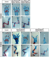
Figure 2. Removal of Bmp2, Bmp4, and Bmp7 from the AER resulted in polydactyly, interdigital webbing, and split hand foot malformations.
Skeleton preparation (A-H) of control and mutant fore- and hindlimbs of newborn mice (P0). Removal of either five of the six Bmp alleles from the forelimb or all known Bmp alleles from the forelimb AER (triple mutants) produced autopod patterning defects (A-C). Removal of five of the six Bmp alleles expressed in the AER (Bmp7 f/+; Bmp2f/f, Bmp4 f/f, Msx2-cre) resulted in the truncation of medial digits in hindlimbs (E and F). Removal of all six Bmp alleles from the AER (Bmp7 f/f; Bmp2f/f, Bmp4 f/f, Msx2-cre) produced severe truncation of medial and/or anterior digits (G and H). In the hindlimbs two examples are shown for each genotype. Truncation of the tibia was observed in 3/10 (30%) of triple mutant hindlimbs (“*” in H’). No other defects in proximal/distal patterning were observed in either the fore- or hindlimbs. A’-H’ are images of the entire limbs.
Figure 3
Figure 3. Removal of Bmp2, Bmp4, and Bmp7 from the AER resulted in an initial broadening and then loss of Fgf8 expression in the AER but normal Fgf10 expression.
In the forelimb, where the Msx2-cre allele instigates recombination after the AER has formed, a slight broadening of Fgf8 expression was observed in the AER (A-C). In E10.5 hindlimb buds containing only a single allele of Bmp7 (H; Bmp7 f/+; Bmp2f/f, Bmp4 f/f, Msx2-cre) or no functional Bmp alleles (I; Bmp7 f/f; Bmp2f/f, Bmp4 f/f, Msx2-cre), Fgf8 expression was broader throughout the AER. In E11.5 limb buds, Fgf8 expression was severely decreased in the central and anterior AER in embryos that contained only a single allele of Bmp7 (E and K; Bmp7 f/+; Bmp2f/f, Bmp4 f/f, Msx2-cre) or no functional Bmp alleles (F and L; Bmp7 f/f; Bmp2f/f, Bmp4 f/f, Msx2-cre). In E10.5 hindlimb buds, Fgf10 expression was not reduced in the limb bud mesenchyme that contained only a single allele of Bmp7 or no functional Bmp alleles. The morphology of the distal limb bud was irregular in hindlimbs that contained only one functional Bmp allele (K, Q) or lacked all Bmp alleles (L, R). No differences in Fgf10 expression were observed in the forelimbs (data not shown).
Figure 4
Figure 4. P63 and CD44 expression were decreased in triple mutants.
(A-D) Immunostaining for ?Np63 on sections of wild type and Bmp triple mutants (Bmp7 f/f; Bmp2f/f, Bmp4 f/f, Msx2-cre). ?Np63 was expressed in the AER and epithelial cells of E10.5 and E11.5 control (A and C) and triple mutant hindlimbs (B and D). In E10.5 triple mutants, the AER was elongated and thinner (B). By E11.5 (D), no visible AER was present. (E-H) Immunostaining for CD44 on sections of wild type (E and G) and triple mutant (F and H; Bmp7 f/f; Bmp2f/f, Bmp4 f/f, Msx2-cre) hindlimbs. CD44 expression was severely decreased in the Bmp triple mutants at E10.5 and E11.5. All sections shown are from the central AER. Scale bar = 20 µm.
Figure 5
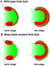
Figure 5. Proposed role for Bmp signaling in the AER.
(A) In wild type limb buds, Bmps are initially expressed in the AER and the anterior and posterior mesenchyme. At later stages of development, Bmp expression is maintained in the AER and in the underlying mesenchyme. (B) Removal of Bmp ligands in the AER resulted in an abnormal expansion (early) and then loss (late) of the central AER. This resulted in defects in autopod patterning but not in limb outgrowth. During early limb bud development, BMP proteins produced in the anterior and posterior limb bud mesenchyme may partially rescue BMP signaling in these regions of the AER (arrows). The central AER appears to require early Bmp expression within this structure. During later development BMP proteins are expressed in the mesenchyme underneath the AER but expression at this time point in not sufficient to maintain a functional AER. Red  =  Bmp expression, green  =  limb bud mesenchyme, white  =  mutant AER.

References

    1. Martin P. Tissue patterning in the developing mouse limb. Int J Dev Biol. 1990;34:323–336. - PubMed
    1. Loomis CA, Kimmel RA, Tong CX, Michaud J, Joyner AL. Analysis of the genetic pathway leading to formation of ectopic apical ectodermal ridges in mouse Engrailed-1 mutant limbs. Development. 1998;125:1137–1148. - PubMed
    1. Guo Q, Loomis C, Joyner AL. Fate map of mouse ventral limb ectoderm and the apical ectodermal ridge. Dev Biol. 2003;264:166–178. - PubMed
    1. Saunders JW. The proximo-distal sequence of the origin of the parts of the chick wing and the role of the ectoderm. Journal of Experimental Zoology. 1948;108:363–403. - PubMed
    1. Dudley AT, Ros MA, Tabin CJ. A re-examination of proximodistal patterning during vertebrate limb development. Nature. 2002;418:539–544. - PubMed

Publication types

MeSH terms

LinkOut - more resources