. 2012;7(5):e37854.
doi: 10.1371/journal.pone.0037854. Epub 2012 May 25.

Down-regulation of GEP100 causes increase in E-cadherin levels and inhibits pancreatic cancer cell invasion

Affiliations

Down-regulation of GEP100 causes increase in E-cadherin levels and inhibits pancreatic cancer cell invasion

Chuan-gao Xie et al. PLoS One. 2012.

Abstract

Aims: Invasion and metastasis are major reasons for pancreatic cancer death and identifying signaling molecules that are specifically used in tumor invasion is of great significance. The purpose of this study was to elucidate the role of GEP100 in pancreatic cancer cell invasion and metastasis and the corresponding molecular mechanism.

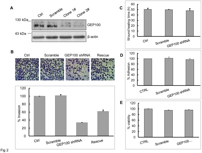
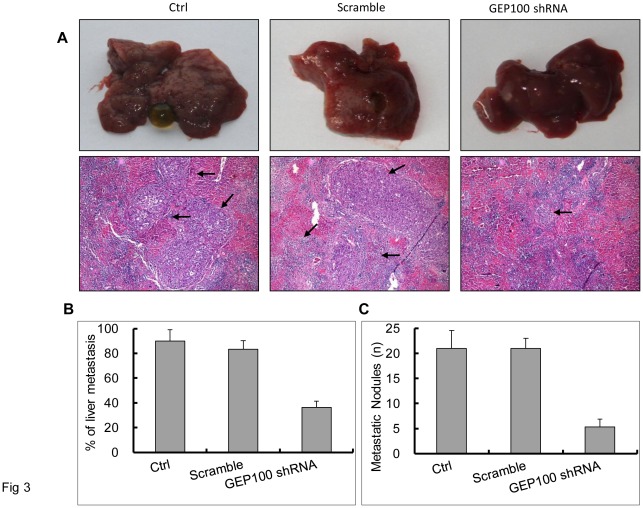
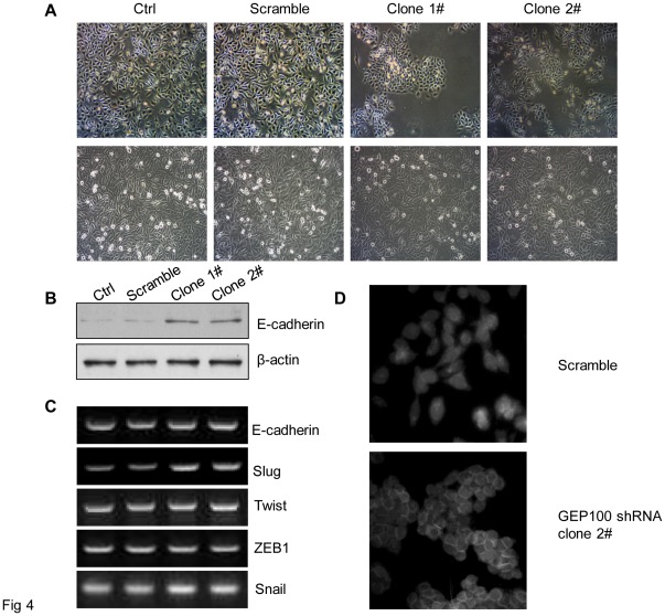
Methods: Stable cell lines with GEP100 knocked-down were established by transfecting GEP100 shRNA vector into PaTu8988 cells and selected by puromycin. qRT-PCR and Western blot were performed to detect gene expression. Matrigel-invasion assay was used to detect cancer cell invasion in vitro. Liver metastasis in vivo was determined by splenic injection of indicated cell lines followed by spleen resection. Immunofluorescence study was used to detect the intracellular localization of E-cadherin.

Results: We found that the expression level of GEP100 protein was closely related to the invasive ability of a panel of 6 different human pancreatic cancer cell lines. Down-regulation of GEP100 in PaTu8988 cells significantly decreased invasive activity by Matrigel invasion assay, without affecting migration, invasion and viability. The inhibited invasive activity was rescued by over-expression of GEP100 cDNA. In vivo study showed that liver metastasis was significantly decreased in the PaTu8988 cells with GEP100 stably knocked-down. In addition, an epithelial-like morphological change, mimicking a mesenchymal to epithelial transition (MET) was induced by GEP100 down-regulation. The expression of E-cadherin protein was increased 2-3 folds accompanied by its redistribution to the cell-cell contacts, while no obvious changes were observed for E-cadherin mRNA. Unexpectedly, the mRNA of Slug was increased by GEP100 knock-down.

Conclusion: These findings provided important evidence that GEP100 plays a significant role in pancreatic cancer invasion through regulating the expression of E-cadherin and the process of MET, indicating the possibility of it becoming a potential therapeutic target against pancreatic cancer.


Conflict of interest statement

Competing Interests: The authors have declared that no competing interests exist.

Figures

Figure 1
Figure 1. GEP100 expression correlates closely with pancreatic cancer cell invasive ability.
(A) Invasive abilities of a panel of 6 pancreatic cancer cell lines were analyzed by Matrigel invasion assay for 24 hours. AsPc-1 and PaTu8988 showed high invasive abilities. (B) Western blot analysis of the expression of GEP100, Arf6, E-cadherin and vimentin protein in different cell lines. AsPc-1 and PaTu8988 showed the strongest expression of GEP100 protein, followed by SW1990, CFPAC-1, Panc-1 and BxPC-3, demonstrating a close relationship between GEP100 expression level and cell invasive ability. No obvious relationship between the expression of Arf6 protein and invasive ability was detected. E-cadherin expression could easily be detected in the low-invasive cell lines, but only a very weak expression was detected in the highly-invasive cell lines. Vimentin was expressed in the highly-invasive cell lines. (C) RT-PCR analysis of the expression of GEP100, Arf6, E-cadherin and vimentin mRNA. The expression of GEP100 mRNA could be detected in all the six cell lines and no obvious correlation with the invasive ability was found. This was the same case for Arf6 and vimentin mRNA. E-cadherin mRNA showed a close association with the invasive ability of different cell lines. β-actin mRNA was used as a control.
Figure 2
Figure 2. Down-regulation of GEP100 inhibits pancreatic cancer cell invasion.
(A) A stable cell line with GEP100 knocked-down was obtained by transfecting psuper-retro-puro-GEP100 into PaTu8988 cells and selected by 1.5 µg/ml of puromycin. Down-regulation of GEP100 was confirmed by Western blot. β-actin was used as a control. (B) Invasion assay showed that GEP100 down-regulation decreased the number of cells penetrated through Matrigel-coated membrane. The data were presented and graphed as percentages calculated by normalizing values obtained for the untreated cells as 100%. GEP100 knock-down decreased the number of cells penetrated through Matrigel to about 35% compared with the control group. Re-expression of GEP100 in shRNA treated cells restored the ability of invasion to about 60%. (C, D, E) GEP100 down-regulation showed no significant effects on cell migration, invasion and viability. Ctrl, control.
Figure 3
Figure 3. GEP100 down-regulation inhibits liver metastasis of pancreatic cancer cells in the Balb/c nude mice with splenic injection.
(A) Macroscopic findings of the resected liver. Stable PaTu8988 cells as indicated were splenicly injected followed by spleen resection. 4 weeks later, livers were removed for observation. Most of the livers from the control and scramble shRNA groups became small and rough, with large whitened areas seen on the surface. For the GEP100 shRNA-injected group, only small spots were found at some of the liver surface. Metastasis was confirmed under a microscope and the sites of metastasis were indicated by arrows (hematoxylin and eosin, x100). (B) Percentages of liver metastasis in the Balb/c nude mice were listed. A statistically significant difference was obtained between control and experimental group (P≤0.05). (C) Average numbers of metastatic nodules in livers were calculated and graphed. Briefly, after a fixation with 10% formalin overnight, the liver was cut into 2 mm slices and 5 sections from approximately the same positions for each liver were estimated under microscope. A statistically significant difference was obtained between control and experimental group (P≤0.05). Ctrl, control.
Figure 4
Figure 4. GEP100 down-regulation induces epithelial change of cancer cells and up-regulation of E-cadherin protein.
(A) A mesenchymal to epithelial change was observed in the cells stably knocked-down for GEP100. The upper and lower panels showed the morphological appearances at a low cell density and when the cells reached confluence, respectively. Comparing with the control and scramble groups, the cell in the experimental group became epithelial-like and adhesive. (B) Western blot showed that the expression level of E-cadherin protein was increased about 3-fold in the experimental group compared with the control group. (C) RT-PCR analysis of E-cadherin mRNA and its transcription regulators. The expression of E-cadherin mRNA was not affected by GEP100 down-regulation. An increase of Slug mRNA was found in the GEP100 knocked-down cells, while there was no change for Twist, ZEB1 and Snail. (D) Immunofluorescence staining of E-cadherin. Comparing with the scramble group, GEP100 down-regulation redistributed the E-cadherin into the cell-cell contacts.

References

    1. Li J, Wientjes MG, Au JL. Pancreatic cancer: pathobiology, treatment options, and drug delivery. AAPS J. 2010;12:223–232. - PMC - PubMed
    1. Jemal A, Siegel R, Ward E, Hao Y, Xu J, et al. Cancer statistics. CA Cancer J Clin. 2009;59:225–249. - PubMed
    1. Li D, Xie K, Wolff R, Abbruzzese JL. Pancreatic cancer. Lancet. 2004;363:1049–1057. - PubMed
    1. Keleg S, Büchler P, Ludwig R, Büchler MW, Friess H. Invasion and metastasis in pancreatic cancer. Mol Cancer. 2003;2:14. - PMC - PubMed
    1. Jones S, Zhang X, Parsons DW, Lin JC, Leary RJ, et al. Core signaling pathways in human pancreatic cancers revealed by global genomic analyses. Science. 2008;321:1801–1806. - PMC - PubMed

Publication types