. 2012;7(5):e37917.
doi: 10.1371/journal.pone.0037917. Epub 2012 May 25.

Induction of neuronal death by microglial AGE-albumin: implications for Alzheimer's disease

Affiliations

Induction of neuronal death by microglial AGE-albumin: implications for Alzheimer's disease

Kyunghee Byun et al. PLoS One. 2012.

Abstract

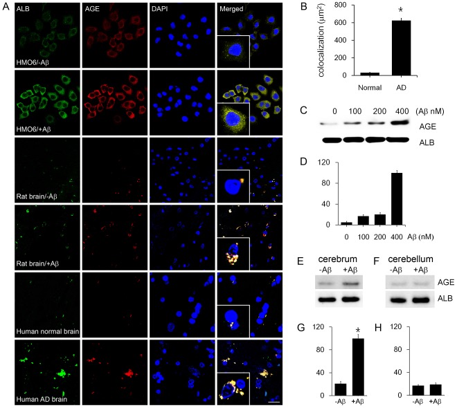
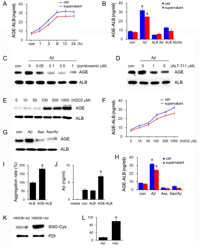
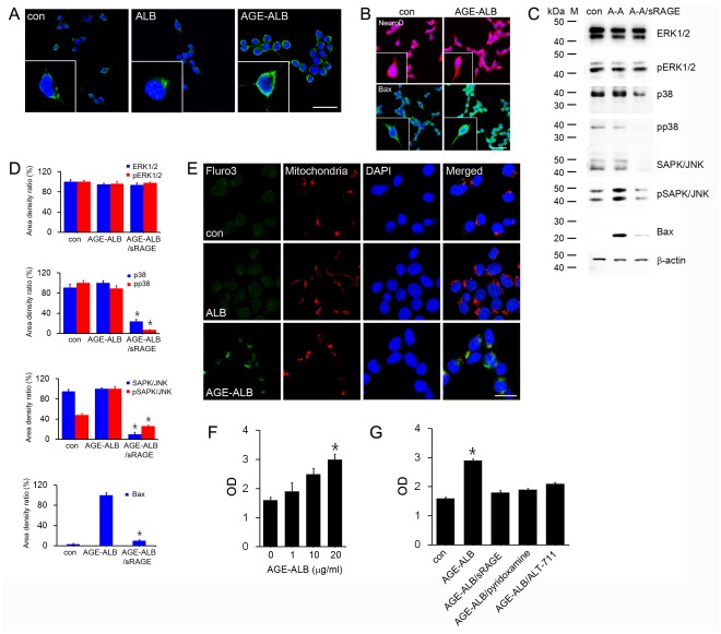
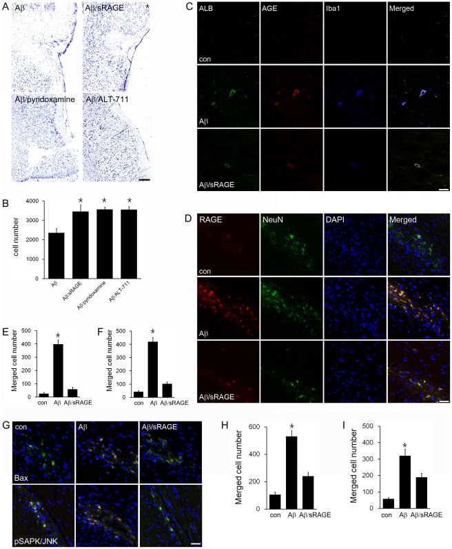
Advanced glycation end products (AGEs) have long been considered as potent molecules promoting neuronal cell death and contributing to neurodegenerative disorders such as Alzheimer's disease (AD). In this study, we demonstrate that AGE-albumin, the most abundant AGE product in human AD brains, is synthesized in activated microglial cells and secreted into the extracellular space. The rate of AGE-albumin synthesis in human microglial cells is markedly increased by amyloid-β exposure and oxidative stress. Exogenous AGE-albumin upregulates the receptor protein for AGE (RAGE) and augments calcium influx, leading to apoptosis of human primary neurons. In animal experiments, soluble RAGE (sRAGE), pyridoxamine or ALT-711 prevented Aβ-induced neuronal death in rat brains. Collectively, these results provide evidence for a new mechanism by which microglial cells promote death of neuronal cells through synthesis and secretion of AGE-albumin, thereby likely contributing to neurodegenerative diseases such as AD.


Conflict of interest statement

Competing Interests: The authors have declared that no competing interests exist.

Figures

Figure 1
Figure 1. Distribution and synthesis of AGE-albumin in microglial cells and rat or human brains.
(A) Triple-labeled confocal microscopic image analyses were used to study the distribution and relative levels of albumin (green), AGE (red) and DAPI (blue) in the HMO6 microglial cells and entorhinal cortex of rat brains before or after Aβ treatment as well as cerebral cortex of human brains from normal or AD individuals. HMO6 cells and rats were treated with Aβ1–42 described in the Materials and Methods section. Scale bar = 50 µm. These results represent similar images of 5 independent analyses. (B) The AGE-albumin positive particles in cortex of human AD brain were significantly different from the normal brains (p<0.05), as determined by densitometric analysis using Zeiss Zen 2009 software. (C, D) The degree of AGE-albumin synthesis in HMO6 cells was determined by immunoblot analysis (C) and densitometric analysis (D) after exposure to different concentrations of Aβ1–42, as indicated. The level of albumin is shown as an internal control for equal protein loading per lane. (E-H) The immunoblots of AGE-albumin in rat cerebrum (E, G) and cerebellum (F, H), with or without Aβ1–42 treatment and densitometric analyses, are shown. *, significantly different (P<0.001) from the level of AGE-albumin without Aβ1–42 treatment.
Figure 2
Figure 2. Synthesis of AGE-albumin in microglial cells but not from astrocytes, oligodendrocytes or neurons from human primary brain cells.
(A) Triple-labeled fluorescent microscopic image analyses were used to demonstrate co-localization of AGE (green), albumin (red), and a specific marker of different cells (blue) in human primary brain cells. Representative images of microglial cells (Iba1), GFAP (an astrocyte marker), Olig2 (an oligodendrocyte marker), and NeuroD (a neuronal marker) in the human primary brain cells are shown. Similar results were observed in 5 independent analyses. Scale bar = 50 µm.
Figure 3
Figure 3. Synthesis of AGE-albumin in human microglial cells and its extracellular secretion.
(A) The time-dependent changes in intracellular (cell lysate) and extracellular (supernatant) AGE-albumin in HMO6 cells, treated with Aβ1–42 for 1, 3, 6, 12, 24 h, were determined by ELISA. (B) The amounts of intracellular (cell lysate) and extracellular (supernatant) AGE-albumin in HMO6 cells, exposed to 3 different conditions as indicated, were determined by ELISA. The microglial cells were treated with: Aβ1–42 alone (5 nM) for 6 h, anti-albumin antibody (ALB Ab, 1 µM) for 24 h, or Aβ1–42 treatment after exposure to anti-albumin antibody overnight. (C-H) The amounts of AGE-albumin were determined by immunoblot analysis of the whole cell lysates of HMO6 cells exposed to different concentrations of pyridoxamine (from 0 to 1 µM) (C), ALT-711 (from 0 to 5 µM) (D), hydrogen peroxide (from 0 to 1,000 µM) (E, F) or 5 µM ascorbic acid (G, H) for 6 h in the absence or presence of 5 nM Aβ1–42 peptide. (I) Relationship between AGE-albumin and Aβ, contributing to Aβ aggregation. Aβ aggregation rates in HMO6 cells, treated with albumin alone or AGE-albumin, were determined by ThT fluorescence analysis. (H) HMO6 cells were exposed to albumin (ALB) or AGE-albumin (AGE-ALB) for 24 h. The respective amounts of Aβ in the culture media from untreated and AGE-albumin-treated cells were measured by ELISA. (I, J) Increased S-nitrosylation of PDI in HMO6 cells after treatment with Aβ. Microglial cells were exposed to Aβ1–42 (400 nM) for 6 h. PDI in whole cell lysates (0.4 mg protein/sample) was immunoprecipitated with the specific antibody. The immunoprecipitated PDI was subjected to immunoblot analysis with anti-S-NO-Cys or anti-PDI antibody. *, Significantly different from control or albumin-treated cells by densitometric analysis (p<0.05).
Figure 4
Figure 4. Induction of neuronal cell death by AGE-albumin through up-regulation of RAGE, mitochondrial calcium influx, and MAPK-Bax pathway.
(A) The relative levels of RAGE (green) or DAPI (blue) in human primary neuronal cells, before or after albumin (ALB) or AGE-albumin (AGE-ALB) treatment for 6 h, were evaluated by double confocal microscopic image analyses. Similar results were observed in 5 independent analyses. (B) Double confocal microscopic images simultaneously show the neuronal marker (NeuroD) and relative levels of Bax (green), or DAPI (blue) in human neuronal cells before or after AGE-ALB treatment for 6 h. (C, D) Whole cell lysates (0.01 mg protein/lane) of human neuronal cells, before or after AGE-albumin exposure, were subjected to immunoblot analysis to determine the levels of ERK1/2, pERK1/2, p38, pp38, pSAPK/JNK, and Bax with specific molecular weight markers (M). β-Actin was used as an internal control for equal protein loading for each lane. (E) Increased level of mitochondrial calcium was evaluated by triple labeled confocal microscopic image analysis before (top panel) and after human neuronal cells were exposed to ALB (middle) or AGE-albumin (bottom): calcium concentration (Fluor-3, green), mitochondria (red), or DAPI-stained nuclei (blue). Scale bar = 50 µm. These results represent similar images of 5 independent analyses. (F, G) The rate of cell death was determined by the apoptosis assay after human neuronal cells were exposed to different concentrations of AGE-albumin alone. (F), or 20 µg/mL AGE-albumin treatment in the absence or presence of co-treatment with sRAGE, pyridoxamine or ALT-711 for 24 h (G).
Figure 5
Figure 5. Protection of Aβ-mediated neuronal cell death by sRAGE, pyridoxamine or ALT-711 through decreasing RAGE levels.
(A, B) The relative levels of neurons in rat brains were evaluated by cresyl violet staining after Aβ injection without or with sRAGE, pyridoxamine or ALT-711 co-treatment for 72 h. *, Significantly increased in Aβ/sRAGE, Aβ/pyridoxamine and Aβ/ALT-711 co-treated samples compared to Aβ treatment only (p<0.01). (C) Triple confocal microscopic images simultaneously show the relative numbers of AGE, albumin, or Iba1 positive cells in the rat entorhinal cortex (EC) before or after Aβ or Aβ/sRAGE injection for 72 h. These results represent similar images of 5 independent analyses. (D) *, The number of the triple-labeled cells (AGE/albumin/Iba-1 positive cells) significantly increased in whole EC area of Aβ injected rat brain but decreased dramatically in Aβ/sRAGE treated rat brain (p<0.01). (E) The levels of RAGE positive neuronal cells were evaluated by triple-labeled confocal microscopic image analysis in entorhinal cortex of control, Aβ, or Aβ/sRAGE injected rat brains. (F) *, The number of RAGE positive neuronal cells significantly increased in Aβ injected rat brain but decreased dramatically in Aβ/sRAGE treated rat brain (p<0.01). (G-I) The number of Bax or pSAPK/JNK positive neurons was evaluated by confocal microscopy. (H, I) *, The number of Bax positive neuronal cells (H) or pSAPK/JNK positive neuronal cells (I) significantly increased in Aβ injected rat brain but decreased dramatically in Aβ/sRAGE treated rat brain (p<0.01). Scale bar = 50 µm.

Similar articles

Cited by

References

    1. Itagaki S, McGeer PL, Akiyama H, Zhu S, Selkoe D. Relationship of microglia and astrocytes to amyloid deposits of Alzheimer’s disease. J Neuroimmunol. 1989;24:173–182. - PubMed
    1. Luber-Narod J, Rogers J. Immune system associated antigens expressed by cells of the human central nervous system. Neurosci Lett. 1988;94:17–22. - PubMed
    1. McGeer PL, Itagaki S, Tago H, McGeer EG. Reactive microglia in patients with senile dementia of the Alzheimer type are positive for the histocompatibility glycoprotein HLA-DR. Neurosci Lett. 1987;79:195–200. - PubMed
    1. Eikelenboom P, Rosemuller AJ, Hoozemans JI, Veerhuis R, van Gool WA. Neuroinflammation and Alzheimer’s disease: Clinical and therapeutic implications. Alzheimer Dis Assoc Disord. 2000;14:S54–61. - PubMed
    1. Neuroinflammation Working Group. Inflammation and Alzheimer’s disease. Neurobiol Aging. 2000;21:383–421. - PMC - PubMed

Publication types

MeSH terms

Substances