. 2012;7(5):e38129.
doi: 10.1371/journal.pone.0038129. Epub 2012 May 25.

Inhibition of neuroblastoma tumor growth by targeted delivery of microRNA-34a using anti-disialoganglioside GD2 coated nanoparticles

Affiliations

Inhibition of neuroblastoma tumor growth by targeted delivery of microRNA-34a using anti-disialoganglioside GD2 coated nanoparticles

Amanda Tivnan et al. PLoS One. 2012.

Abstract

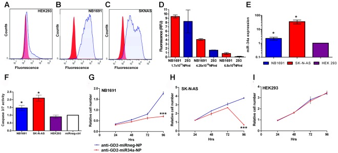
Background: Neuroblastoma is one of the most challenging malignancies of childhood, being associated with the highest death rate in paediatric oncology, underlining the need for novel therapeutic approaches. Typically, patients with high risk disease undergo an initial remission in response to treatment, followed by disease recurrence that has become refractory to further treatment. Here, we demonstrate the first silica nanoparticle-based targeted delivery of a tumor suppressive, pro-apoptotic microRNA, miR-34a, to neuroblastoma tumors in a murine orthotopic xenograft model. These tumors express high levels of the cell surface antigen disialoganglioside GD2 (GD(2)), providing a target for tumor-specific delivery.

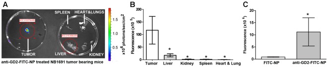
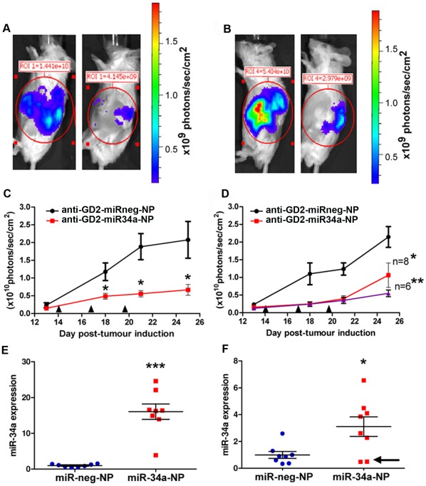
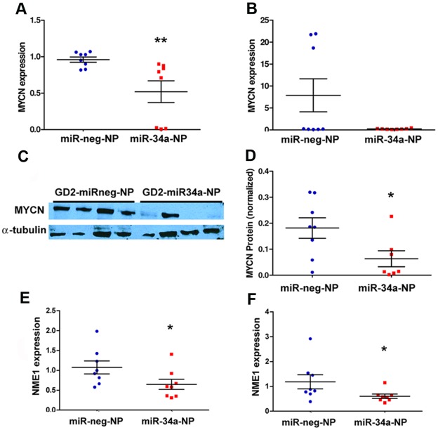
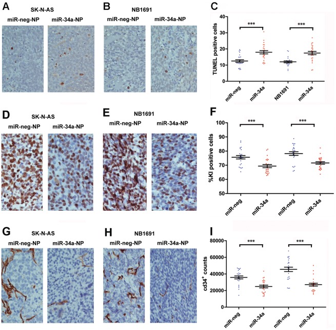
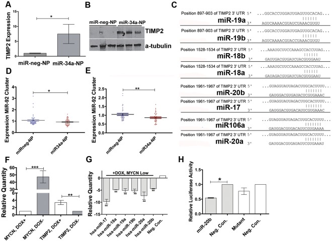
Principal findings: Nanoparticles encapsulating miR-34a and conjugated to a GD(2) antibody facilitated tumor-specific delivery following systemic administration into tumor bearing mice, resulted in significantly decreased tumor growth, increased apoptosis and a reduction in vascularisation. We further demonstrate a novel, multi-step molecular mechanism by which miR-34a leads to increased levels of the tissue inhibitor metallopeptidase 2 precursor (TIMP2) protein, accounting for the highly reduced vascularisation noted in miR-34a-treated tumors.

Significance: These novel findings highlight the potential of anti-GD(2)-nanoparticle-mediated targeted delivery of miR-34a for both the treatment of GD(2)-expressing tumors, and as a basic discovery tool for elucidating biological effects of novel miRNAs on tumor growth.


Conflict of interest statement

Competing Interests: The authors have declared that no competing interests exist.

Figures

Figure 1
Figure 1. Specific uptake of anti-GD2-nanoparticles exclusively by GD2 expressing cells.
Effects on growth and caspase activity by anti-GD2 conjugated nanoparticles containing miR-34a. (A) FACS analysis, using an anti-GD2 primary antibody and goat anti-mouse IgG2a-PE secondary antibody indicated little, if any, GD2 surface antigen on HEK293 cells. Conversely NB1691 (B) and SK-N-AS (C) cells showed significant GD2 reactivity. (D) Varying concentrations of FITC-anti-GD2-NPs were added to cell culture media of NB1691 or HEK293 (1×10⁶ cells). A concentration of 6.8×10⁹ particles/ml (40 µg/ml) was the optimal dosage tested in vitro for specific delivery of encapsulated FITC fluorophore to NB1691 neuroblastoma cells with minimal incorporation into HEK293 cells. (E) Anti-GD2-miR34a or control-anti-GD2-nanoparticles were added to adhered neuroblastoma SK-N-AS and NB1691, and to HEK293 cells for 4 hours in standard media. Media was removed, the cells washed and media was replaced. RNA was isolated from these cells after 24 hours and assessed for miR34a levels. Anti-GD2-miR34a-NPs treatment (40 µg/ml) led to ∼2 fold increase in miR-34a levels in NB1691 cells, ∼30 fold increase in SK-N-AS cells (*p<0.05, n=3), with no significant change in miR-34a transcript levels in HEK293 cells. (F) NB1691 and SK-N-AS cells showed a significant increase in caspase 3/7 activity relative to anti-GD2-miRneg-NP-treated cells after 72 hrs (*p<0.05, n=3) while HEK293 cells showed no significant increase in caspase activity under these conditions. Acid phosphatase assays indicated a significant reduction in viable cell numbers for (G) NB1691 and (H) SK-N-AS cells treated with anti-GD2-miR34a-NPs, over a 96 hour period (***p<0.001) relative to anti-GD2-miRneg-NP-treated controls. (I) HEK293 cells showed no significant reduction of viable cells following treatment with anti-GD2-miR34a-NP (p>0.05, n=3).
Figure 2
Figure 2. Targeting of neuroblastoma by systemically administered anti-GD2 conjugated nanoparticles.
(A) Systemic administration through lateral tail injection of FITC-anti-GD2-NPs (1 mg/ml) resulted in targeted delivery of FITC dye predominantly to tumors (indicated by fluorescence intensity color map generated from scans using IVIS instrumentation). (B) FITC-anti-GD2-NP-treated organs have significantly less fluorescence than isolated tumors (*p<0.05, n=4−5) and (C) FITC-anti-GD2-NP treated tumors showed significantly greater presence of FITC dye relative to FITC-NP treated tumors (*p<0.05, n=4−5).
Figure 3
Figure 3. Anti-neuroblastoma effect of anti-GD2 conjugated nanoparticles bearing miR-34a in vivo.
Bioluminescent images representative of mice bearing (A) NB1691luc tumors treated with anti-GD2-miRneg-NP (left) or anti-GD2-miR34a-NP (right), along with representative mice bearing (B) SK-N-ASluc tumors treated with anti-GD2-miRneg-NP (left) or anti-GD2-miR34a-NP (right) were obtained at day 25. Tumor growth curves from (C) mice bearing NB1691luc tumors treated with anti-GD2-miRneg-NP (black line) or anti-GD2-miR34a-NP (red line) and (D) mice bearing SK-N-ASluc tumors treated with anti-GD2-miRneg-NP (black line) or anti-GD2-miR34a-NP (red line). Time points for systemic administration of nanoparticles are indicated by the symbol ▴. Differences in tumor growth between mice injected with anti-GD2-miR34a-NP versus anti-GD2-miRneg-NP were statistically significant for both models (NB1691luc n=8, *p<0.05) (SK-N-ASluc *p<0.05 for n=8 and **p<0.01 for n=6). For the SK-N-ASluc model, two tumors grew significantly faster than the median and are represented in the growth curve for all 8 tumors (red line). The purple line represents the growth curve without these two tumors (n=6). Mature miR-34a transcript levels were significantly higher in anti-GD2-miR34a-NP treated tumours relative to anti-GD2-miRneg-NP-treated control tumors in both the (E) NB1691luc (***p<0.001, n=8) and (F) SK-N-ASluc (*p<0.05, n=8). Notably, the two SK-N-AS tumors with increased growth from (D) had poor uptake of miR-34a (arrow).
Figure 4
Figure 4. Targeting of MYCN by miR-34a in vivo.
(A) MYCN, a validated target of miR-34a, mRNA levels were significantly reduced in GD2-miR34a-NP treated NB1691luc tumors relative to negative controls (**p<0.001, n=8) (B) qPCR analysis of SK-N-ASluc tumors did not indicate a statistically significant difference between anti-GD2-miR34a-NP and anti-GD2-miRneg-NP treated tumours, potentially because levels of MYCN expression are very low in this cell line. Three SK-N-AS tumors treated with negative control nanoparticles appeared to have somewhat higher expression then the median, although this level of expression is very low compared to MYCN amplified tumors (C) MYCN reduction in NB1691luc was validated at a protein level by Western Blot and (D) protein suppression was quantified using densitometry. NME1, a validated MYCN target, had reduced mRNA levels in anti-GD2-miR34a-NP treated NB1691luc tumors (E) and SK-N-ASluc tumors (F) relative to negative controls (*p<0.05, n=8), as evaluated by qPCR.
Figure 5
Figure 5. Pro-apoptotic and anti-angiogenic activity of GD2 targeted nanoparticles containing miR-34a.
Tumors of mice treated with anti-GD2-miR-34a-NPs and negative controls were analyzed by TUNEL staining on paraffin embedded section of SK-N-ASluc (a) and NB1691luc (b) tumors. In both tumor subtypes, treatment with anti-GD2-miR34a-NPs led to a significant increase in apoptosis (c, ***p<0.001, n=8). Immunohistochemistry staining with CD34 showed a marked decrease in the endothelial cell marker subsequent to anti-GD2-miR34a-NP treatment (d-f, ***p<0.001, n=8). KI67 staining showed reduced proliferation in anti-GD2-miR34a-NP treated cohorts (g-i, ***p<0.001, n=8).
Figure 6
Figure 6. Molecular events involved with miR-34a anti-angiogenic effects.
(A) qPCR and (B) western blot analyses of TIMP2 mRNA (n=4 tumors) and protein levels (n=3 tumors) from NB1691 tumors treated with anti-GD-miR-neg-NP or anti-GD20miR34a-NP. (C) Computationally predicted target sites on the 3′ UTR of TIMP2 for miR-92 polycistronic cluster members from chromosome 13 and X. qPCR analysis of miR-92 cluster members from chromosome 13 (D) and chromosome X (E) in tumors treated with anti-GD-miR-neg-NP (n=8) or anti-GD20miR34a-NP (n=8). (F) qPCR analysis of MYCN and TIMP2 expression in SHEP TET21 cells possessing a repressible MYCN transgene. Expression levels of these genes in cells treated (DOX+) and untreated (DOX-) with doxycycline are displayed. (G) Expression levels of miR-92 cluster members in SHEP TET21 following treatment with doxycycline (MYCN low expression state)(n=5 biological replicate experiments). (H) Luciferase assays following co-transfection of NB1691 cells with a reporter construct containing a wild type TIMP2 3′ UTR region and either miR-20b mimics or a negative control oligonucleotide (n=2). Luciferase activity following co-transfection of the same cell line with a plasmid having a mutated binding site for miR-20b and either miR-20b mimics or a negative control oligonucleotide is also displayed.

References

    1. Wagner LM, Danks MK. New therapeutic targets for the treatment of high-risk neuroblastoma. J Cell Biochem. 2009;107:46–57. - PubMed
    1. Ritter G, Livingston PO. Ganglioside antigens expressed by human cancer cells. Semin Cancer Biol. 1991;2:401–409. - PubMed
    1. Handgretinger R, Anderson K, Lang P, Dopfer R, Klingebiel T, et al. A phase I study of human/mouse chimeric antiganglioside GD2 antibody ch14.18 in patients with neuroblastoma. Eur J Cancer. 1995;31A:261–267. - PubMed
    1. Yu AL, Uttenreuther-Fischer MM, Huang CS, Tsui CC, Gillies SD, et al. Phase I trial of a human-mouse chimeric anti-disialoganglioside monoclonal antibody ch14.18 in patients with refractory neuroblastoma and osteosarcoma. J Clin Oncol. 1998;16:2169–2180. - PubMed
    1. Zeytin HE, Tripathi PK, Bhattacharya-Chatterjee M, Foon KA, Chatterjee SK. Construction and characterization of DNA vaccines encoding the single-chain variable fragment of the anti-idiotype antibody 1A7 mimicking the tumor-associated antigen disialoganglioside GD2. Cancer Gene Ther. 2000;7:1426–1436. - PubMed

Publication types

MeSH terms