. 2012 Feb 16;3(4):543-554.
doi: 10.1021/jz2014117. Epub 2012 Feb 8.

Proton Coupled Electron Transfer and Redox Active Tyrosines: Structure and Function of the Tyrosyl Radicals in Ribonucleotide Reductase and Photosystem II

Affiliations

Proton Coupled Electron Transfer and Redox Active Tyrosines: Structure and Function of the Tyrosyl Radicals in Ribonucleotide Reductase and Photosystem II

Bridgette A Barry et al. J Phys Chem Lett. .

Abstract

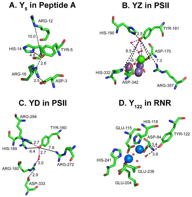
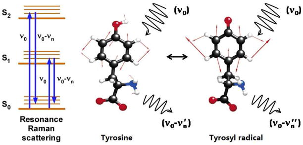
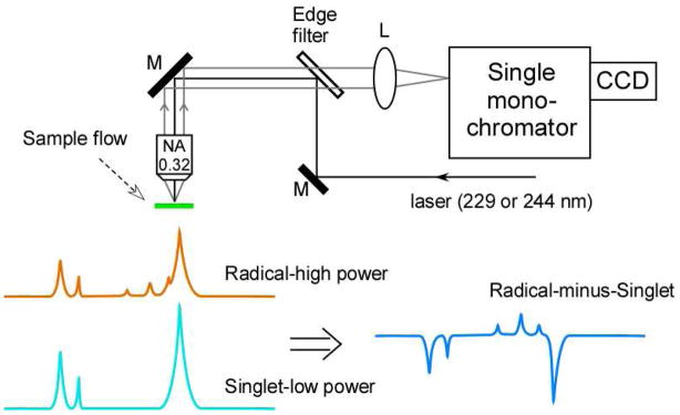
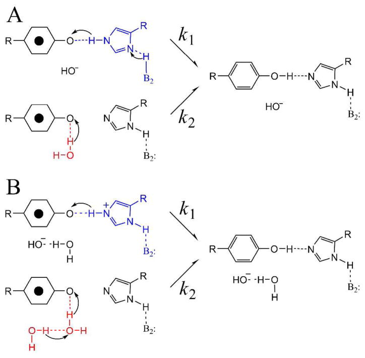
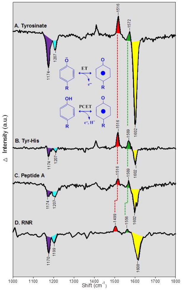
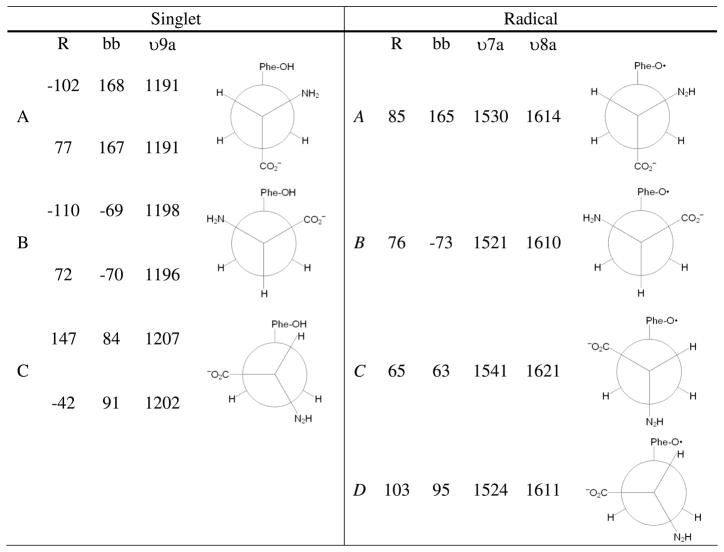
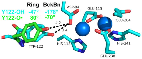
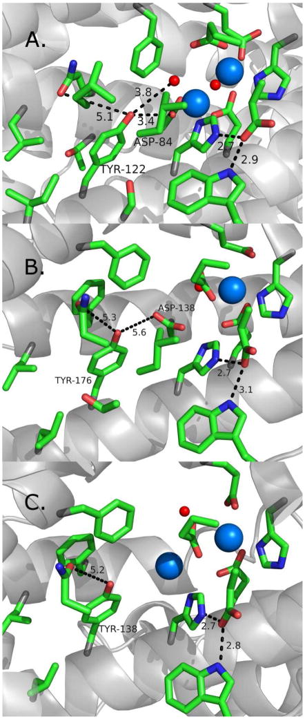
Proton coupled electron transfer (PCET) reactions are important in many biological processes. Tyrosine oxidation/reduction can play a critical role in facilitating these reactions. Two examples are photosystem II (PSII) and ribonucleotide reductase (RNR). RNR is essential in DNA synthesis in all organisms. In E. coli RNR, a tyrosyl radical, Y122(•), is required as a radical initiator. Photosystem II (PSII) generates molecular oxygen from water. In PSII, an essential tyrosyl radical, YZ(•), oxidizes the oxygen evolving center. However, the mechanisms, by which the extraordinary oxidizing power of the tyrosyl radical is controlled, are not well understood. This is due to the difficulty in acquiring high-resolution structural information about the radical state. Spectroscopic approaches, such as EPR and UV resonance Raman (UVRR), can give new information. Here, we discuss EPR studies of PCET and the PSII YZ radical. We also present UVRR results, which support the conclusion that Y122 undergoes an alteration in ring and backbone dihedral angle when it is oxidized. This conformational change results in a loss of hydrogen bonding to the phenolic oxygen. Our analysis suggests that access of water is an important factor in determining tyrosyl radical lifetime and function. TOC graphic.


Figures

Figure 1
Figure 1
Thermodynamic cycle, illustrating the linkage of pKa and midpoint potential, for tyrosine oxidation. The concerted pathway for proton and electron transfer is labeled as CPET.
Figure 2
Figure 2
Comparison of the hydrogen bonding environments of four redox active tyrosines. Shown in (A) Y5 in biomimetic beta hairpin (peptide A), in (B) YZ in PSII, in (C) YD in PSII, and in (D) Y122, in the beta subunit of E. coli RNR. Predicted hydrogen bonds are shown in orange dashed lines, distances are given in black dashed lines. In B, the manganese ions are purple, and the calcium ion is green. In D, the iron ions are blue. Oxygen atoms are shown as red spheres in B–D.
Figure 3
Figure 3
Schematic of the Raman scattering event and of ring stretching (Y8a) vibrational modes, occurring in tyrosine and tyrosyl radical.
Figure 4
Figure 4
Schematic of UV resonance Raman spectrometer and the vibrational difference method used to study redox active tyrosines. A spectrum acquired at low UV probe power, representing the singlet (light blue), is subtracted from a spectrum acquired at high power (orange), to yield the difference spectrum: radical-minus-singlet (dark blue).
Figure 5
Figure 5
Two possible multi-proton pathways (A and B) for PCET and YD. Pathway B involves a hydrogen bonded chain of water molecules, of which two are shown. This model with two competing pathways, one involving more than three protons, was predicted by a proton inventory experiment at high pH. Reprinted from ref .
Figure 6
Figure 6
UV resonance Raman spectra of tyrosyl radicals in (A) tyrosinate, (B) a tyrosinate-histidine dipeptide, (C) peptide A, and (D) ribonucleotide reductase (RNR). The data are difference spectra reflecting: radical-minus-singlet, plotted against the Raman shift in cm−1. The spectra in (A–C) correspond to the ET reaction shown. These data were obtained with a 244 nm probe beam, and the radical was photoinduced by increasing the UV power. The difference spectra were acquired by subtracting a low power scan (340μW, 16 scans, 240s/scan) from the high power scan (3.4 mW, 8 scans, 120 s/scan). The amplitude of low intensity scan was multiplied by a factor of 5 before subtraction. The samples were suspended in a 2H2O buffer containing 5 mM sodium borate, p2H 11. In (D), the spectrum corresponds to the PCET reaction shown. The RNR difference spectrum was obtained chemically, by subtracting data obtained from a met-beta (hydroxyurea-treated) sample, which lacks Y122, from data obtain on a control (untreated) beta sample, which contains Y122. The data were acquired at pH 7.6 with 229 nm excitation (7 scans, 180s/scan). See ref for more experimental details.
Figure 7
Figure 7
Ring {r} and backbone {bb} dihedral angles, plus Y9a (singlet), Y7a (radical), and Y8a (radical) vibrational frequencies, for the lowest energy conformers of tyrosinate and tyrosyl radical.
Figure 8
Figure 8
Structure of singlet Y122 (blue) and predicted structure of Y122 radical (green) based on the observed CO and ring stretching frequency, which are consistent with the B conformer. The conformational change from the B conformer (radical, green) to A conformer (singlet, blue) is predicted to rotate Y122 (singlet) into hydrogen bonding distance with Asp 84 (D84). The blue spheres represent the iron cluster. Distances between D84 and Y122 are in dotted lines and in Angstroms. Ring and backbone (Bckbn) angles for the radical (green) and singlet (blue) are shown.
Figure 9
Figure 9
Comparison of the environments, near the redox active tyrosines, in (A) E. coli, (B) human, and (C) human p53-induced RNR (beta subunit). The structures of the singlet state are shown. The blue and red spheres represent iron and water/oxygen. Distances are in black dotted lines and in Angstroms. The resolutions of the solved X-ray structures are (A) 1.4 Å (B) 2.8 Å and (C) 2.8 Å.

References

    1. Stubbe J. Ribonucleotide Reductases: Amazing and Confusing. J Biol Chem. 1990;265:5329–5332. - PubMed
    1. Barry BA, Babcock GT. Tyrosine Radicals are Involved in the Photosynthetic Oxygen-Evolving system. Proc Nat Acad Sci. 1987;84:7099–7103. - PMC - PubMed
    1. Boerner RJ, Barry BA. Isotopic Labeling and EPR Spectroscopy Show that a Tyrosine Residue is the Terminal Electron Donor, Z, in Manganese-depleted Photosystem II Preparations. J Biol Chem. 1993;268:17151–17154. - PubMed
    1. Kulmacz RJ, Ren Y, Tsai AL, Palmer G. Prostaglandin H synthase: Spectroscopic Studies of the Interaction with Hydroperoxides and with Indomethacin. Biochemistry. 1990;29:8760–8771. - PubMed
    1. Whittaker MM, Whittaker JW. Tyrosine-derived Free Radical in Apogalactose Oxidase. J Biol Chem. 1990;265:9610–9613. - PubMed