

Sex hormones and the QT interval: a review
- PMID: 22663191
- PMCID: PMC3430484
- DOI: 10.1089/jwh.2011.3444
Sex hormones and the QT interval: a review
Abstract
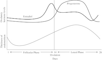
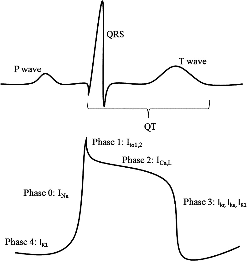
A prolonged QT interval is a marker for an increased risk of ventricular tachyarrhythmias. Both endogenous and exogenous sex hormones have been shown to affect the QT interval. Endogenous testosterone and progesterone shorten the action potential, and estrogen lengthens the QT interval. During a single menstrual cycle, progesterone levels, but not estrogen levels, have the dominant effect on ventricular repolarization in women. Studies of menopausal hormone therapy (MHT) in the form of estrogen-alone therapy (ET) and estrogen plus progesterone therapy (EPT) have suggested a counterbalancing effect of exogenous estrogen and progesterone on the QT. Specifically, ET lengthens the QT, whereas EPT has no effect. To date, there are no studies on oral contraception (OC) and the QT interval, and future research is needed. This review outlines the current literature on sex hormones and QT interval, including the endogenous effects of estrogen, progesterone, and testosterone and the exogenous effects of estrogen and progesterone therapy in the forms of MHT and hormone contraception. Further, we review the potential mechanisms and pathophysiology of sex hormones on the QT interval.
Figures
References
-
- Rautaharju PM. Surawicz B. Gettes LS, et al. AHA/ACCF/HRS recommendations for the standardization and interpretation of the electrocardiogram: Part iv: The ST segment, T and U waves, and the QT interval: A scientific statement from the American Heart Association Electrocardiography and Arrhythmias Committee, Council on Clinical Cardiology; the American College of Cardiology Foundation; and the Heart Rhythm Society: Endorsed by the International Society for Computerized Electrocardiology. Circulation. 2009;119:e241–250. - PubMed
-
- Algra A. Tijssen JG. Roelandt JR. Pool J. Lubsen J. QTc prolongation measured by standard 12-lead electrocardiography is an independent risk factor for sudden death due to cardiac arrest. Circulation. 1991;83:1888–1894. - PubMed
-
- Makkar RR. Fromm BS. Steinman RT. Meissner MD. Lehmann MH. Female gender as a risk factor for torsades de pointes associated with cardiovascular drugs. JAMA. 1993;270:2590–2597. - PubMed
-
- Kannel WB. Wilson PW. D'Agostino RB. Cobb J. Sudden coronary death in women. Am Heart J. 1998;136:205–212. - PubMed
-
- Albert CM. Chae CU. Grodstein F, et al. Prospective study of sudden cardiac death among women in the United States. Circulation. 2003;107:2096–2101. - PubMed
Publication types
MeSH terms
Substances
Grants and funding
LinkOut - more resources
Full Text Sources
Other Literature Sources
