

Controlled Clinical Trial
doi: 10.1016/S0019-4832(12)60082-0.
Diagnostic accuracy of 64-slice multidetector computed tomography in evaluation of post-coronary artery bypass grafts in correlation with invasive coronary angiography
Affiliations
- PMID: 22664806
- PMCID: PMC3860957
- DOI: 10.1016/S0019-4832(12)60082-0
Item in Clipboard
Controlled Clinical Trial
Diagnostic accuracy of 64-slice multidetector computed tomography in evaluation of post-coronary artery bypass grafts in correlation with invasive coronary angiography
Indian Heart J.
2012 May-Jun.
Abstract
64-slice multidetector computed tomography (MDCT) allows more reliable and non-invasive evaluation of the coronary artery bypass grafts for occlusion or stenosis both in symptomatic and asymptomatic patients and also progression of disease in native coronary vessels.
Copyright © 2012 Cardiological Society of India. Published by Elsevier B.V. All rights reserved.
Figures

VR images showing patent sequential arterial graft. D1: diagonal 1, ILA: intra-operative linear array, LAD: left anterior descending artery, LIMA: left internal mammary artery.

VR images showing patent arterial grafts. LAD: left anterior descending artery, LIMA: left internal mammary artery, RIMA: right internal mammary artery, OM: obtuse marginal.

(A) VR image, (B) invasive coronary angiography image showing occluded venous graft.

Maximum intensity projection images showing patent SVGs to native arteries. LAD: left anterior descending artery, SVG: saphenous venous graft, OM: obtuse marginal, LCX: left circumflex, PDA: posterior descending artery.

VR image (A) showing occluded SVG and RA grafts as confirmed with invasive coronary angiography (B,C). RA: radial artery, SVG: saphenous venous graft.
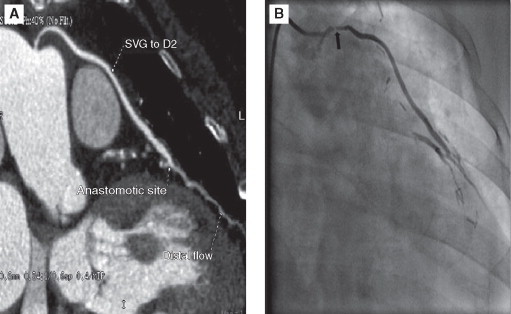
(A) MIP image shows mild stenosis in the body of SVG to D2 graft in comparision with (B) invasive coronary angiography image showing severe stenosis. D2: diagonal 2, MIP: maximum intensity projection, SVG: saphenous venous graft.

Graph age since surgery.
Comment in
-
Diagnostic accuracy of coronary computed tomography angiography in patients post-coronary artery bypass grafting.Indian Heart J. 2012 May-Jun;64(3):261-2. doi: 10.1016/S0019-4832(12)60083-2. Indian Heart J. 2012. PMID: 22664807 Free PMC article. No abstract available.
References
-
- Yusuf S, Reddy S, Ounpuu S, Anand S. Global burden of cardiovascular disease: Part II. Variations in cardiovascular disease by specific ethnic groups and geographic regions and prevention strategies. Circulation. 2001;104:2855–2864. - PubMed
-
- Gupta R. Epidemiological evolution and rise of coronary heart disease in India. South Asian J Prev Cardiol. 1997;1:14–20.
-
- Austen WG, Edwards JE, Frye RL. A reporting system on patients evaluated for coronary artery disease. Report of the Ad Hoc Committee for Grading of Coronary Artery Disease, Council on Cardiovascular Surgery, American Heart Association. Circulation. 1975;51(Suppl 4):5–40. - PubMed
-
- Frazier AA, Qureshi F, Read KM, Gilkeson RC, Poston RS, White CS. Coronary artery bypass grafts: assessment with multidetector CT in the early and late postoperative settings. Radiographics. 2005;25:881–896. - PubMed
-
- Jones CM, Chin KY, Yang GZ. Coronary artery bypass graft imaging with 64-slice multislice computed tomography: literature review. Semin Ultrasound CT MRI. 2008;29:204–213. - PubMed
Publication types
MeSH terms
LinkOut - more resources
Full Text Sources
Medical
