. 2012 Oct;22(10):1453-66.
doi: 10.1038/cr.2012.88. Epub 2012 Jun 5.

CFTR mediates bicarbonate-dependent activation of miR-125b in preimplantation embryo development

Affiliations

CFTR mediates bicarbonate-dependent activation of miR-125b in preimplantation embryo development

Yong Chao Lu et al. Cell Res. 2012 Oct.

Abstract

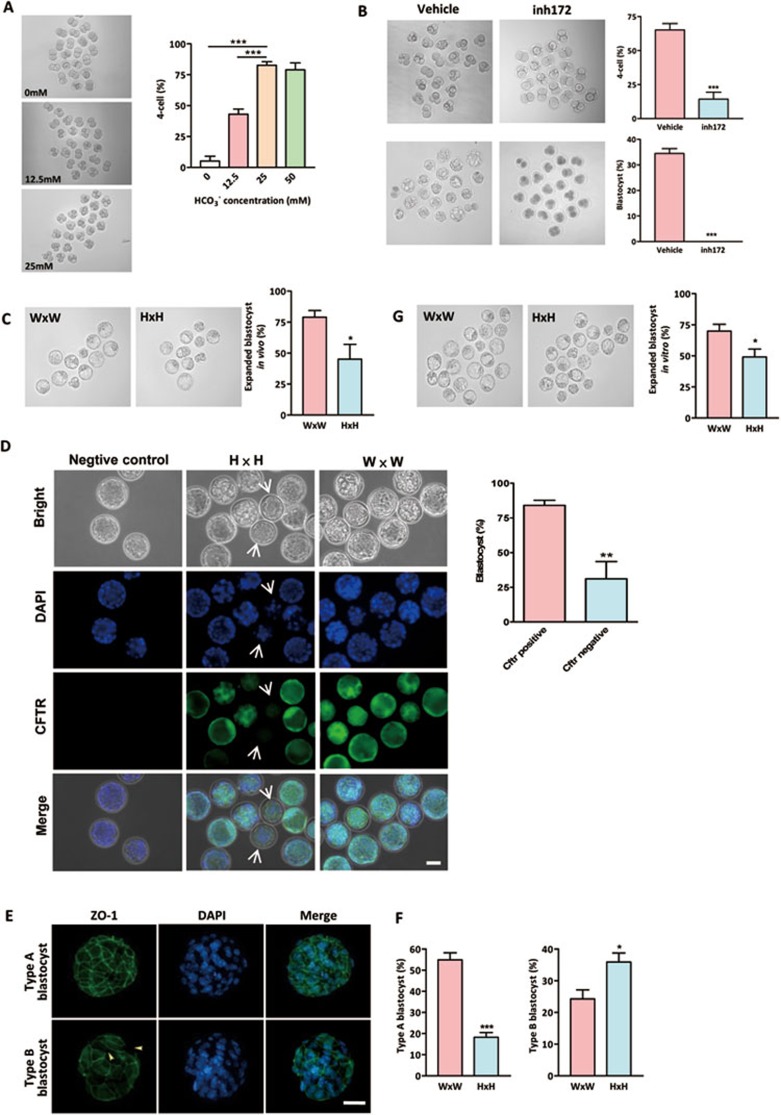
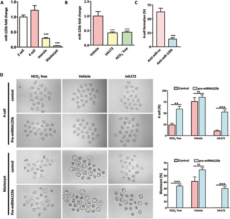
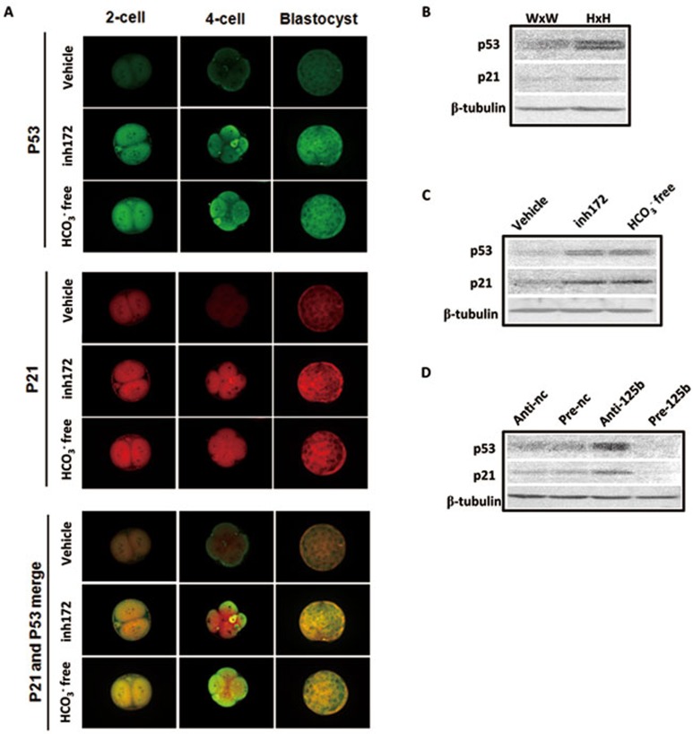
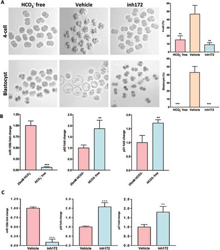
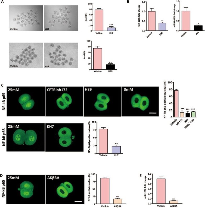
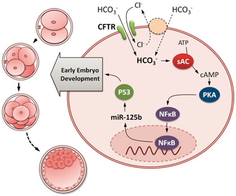
Although HCO(3)(-) is known to be required for early embryo development, its exact role remains elusive. Here we report that HCO(3)(-) acts as an environmental cue in regulating miR-125b expression through CFTR-mediated influx during preimplantation embryo development. The results show that the effect of HCO(3)(-) on preimplantation embryo development can be suppressed by interfering the function of a HCO(3)(-)-conducting channel, CFTR, by a specific inhibitor or gene knockout. Removal of extracellular HCO(3)(-) or inhibition of CFTR reduces miR-125b expression in 2 cell-stage mouse embryos. Knockdown of miR-125b mimics the effect of HCO(3)(-) removal and CFTR inhibition, while injection of miR-125b precursor reverses it. Downregulation of miR-125b upregulates p53 cascade in both human and mouse embryos. The activation of miR-125b is shown to be mediated by sAC/PKA-dependent nuclear shuttling of NF-κB. These results have revealed a critical role of CFTR in signal transduction linking the environmental HCO(3)(-) to activation of miR-125b during preimplantation embryo development and indicated the importance of ion channels in regulation of miRNAs.


Figures

Figure 1
Figure 1
Involvement of CFTR in HCO3-dependent preimplantation embryo development. (A) Effects of HCO3 on preimplantation embryo development. Embryos were cultured in different concentrations of HCO3. Transition from two-cell to four-cell embryos were significantly inhibited by removing (0 mM) or reducing extracellular (12.5 mM) HCO3 (0 mM group: 6/108 embryos; 12.5 mM group: 46/106 embryos; 25 mM group: 95/110 embryos; 50 mM group: 83/105 embryos). (B) CFTRinh172 (inh172) significantly reduced four-cell (n = 4, 12/85 embryos) and blastocyst formation (0/85 embryos) in embryo culture containing 25 mM HCO3 compared to the DMSO-treated vehicle control (four-cell: 57/88 embryos; blastocyst: 30/88). (C) Embryos obtained from Cftr+/− mice (H × H) on 3.5 dpc have reduced percentage of expanded blastocysts (31/68 embryos) as compared to those obtained from Cftr+/+ mice (W × W) (60/79 embryos). (D) Cftr−/− embryos, as indicated by lacking CFTR immunoreactivity (arrow), exhibit a remarkable decrease in blastocyst formation rate (5/13 embryos) as compared to CFTR-positive embryos (38/48 embryos). (E) Embryos were categorized into two types according to the developmental stages and ZO-1 fluorescence: type A blastocysts showed continuous and well-organized ZO-1 expression only at the cell junction, while type B blastocyst showed disrupted expression of ZO-1 at the cell junction (arrow head), and diffused localization in cytoplasm. (F) Embryos from Cftr+/+ and Cftr+/− mice were classified by ZO-1 expression patterns (green) in blastocyst stages. Nuclei were counterstained by DAPI. Embryos obtained from Cftr+/− mice have a significantly reduced percentage of type A blastocyst. (G) Embryos obtained from Cftr+/− mice (H × H) on 1.5 dpc have reduced expanded blastocyst formation (60/122 embryos) after in vitro culture for 72 h as compared to those obtained form Cftr+/+ mice (W × W) (87/126 embryos). Data are presented as mean ± SEM; one-way ANOVA (A); t-test (B-E); *P < 0.05 and ***P < 0.001. Scale bar, 50 μm.
Figure 2
Figure 2
Involvement of miR-125b in CFTR-regulated embryo development. (A) Real-time PCR results showing high miR-125b expression in two-cell and four-cell embryos, but significantly reduced in morula and blastocyst stages (n = 4, 100 embryos/group). (B) MiR-125b expression in two-cell embryo was inhibited by CFTRinh172 treatment (compared to DMSO-treated vehicle control) and in HCO3-free condition (n = 4, 100 embryos/group). (C) Knockdown of miR-125b inhibited four-cell embryo formation (n = 4, 80 embryos/group). (D) Impaired four-cell and blastocyst formation by removal of HCO3 and CFTR inhibition was rescued by injection of miR-125b precursor (pre-miR-125b) (n = 3); HCO3 free + pre-miR-nc group: four-cell (15/62 embryos), blastocyst (0/62 embryos); HCO3 free + pre-miR-125b group: four-cell (36/61 embryos), blastocyst (21/60 embryos); Vehicle + pre-miR-nc group: four-cell (46/60 embryos), blastocyst (25/60 embryos); Vehicle + pre-miR-125b group: four-cell (55/64 embryos), blastocyst (38/64 embryos); CFTRinh172 + pre-miR-nc group: four-cell (10/60 embryos), blastocyst (0/60 embryos); CFTRinh172 + pre-miR-125b group: four-cell (33/63 embryos), blastocyst (19/63 embryos). Data are presented as mean ± SEM; One way ANOVA (A, B); t-test (C, D); **P < 0.01, ***P < 0.001, ns – not significant. Scale bar, 50 μm.
Figure 3
Figure 3
p53 and p21 are the downstream targets of CFTR-HCO3-miR-125b pathway. (A) Immunofluorescent results showing increased expression of p53 (green) and p21 (red) in HCO3-free condition or after CFTRinh172 treatment (5-10 embryos/group). (B) Western blot results showing increased expression of p53 and p21 in two-cell embryos from Cftr+/− mice (H × H) compared to those from Cftr+/+ (W × W) mice. (C) Western blot results showing upregulation of p53 and p21 by CFTRinh172 or HCO3-free condition in two-cell embryos. (D) Western blot results showing that transfection of anti-miR-125b oligo leads to increased expression of p53 and p21, while transfection of miR-125b precursor leads to decreased expression of p53 and p21 in two-cell embryos. Scale bar, 50 μm.
Figure 4
Figure 4
Involvement of CFTR and HCO3 in human embryo development and regulation of miR-125b-p53 cascade. (A) Decreased four-cell and blastocyst formation in HCO3-free condition or after CFTRinh172 (inh172) treatment (n = 3; HCO3-free group: four-cell (9/60 embryos), blasocyst (0/60 embryos); Vehicle group: four-cell (30/63 embryos), blastocyst (27/63 embryos); CFTRinh172 group: four-cell (6/62 embryos), blastocyst (0/62) ). (B, C) Real-time PCR showed expression of miR-125b, p53 and p21 under HCO3-free condition (B) or after CFTRinh172 treatment (C) (n = 4 100 embryos/group). Inhibition of CFTR or cultured in HCO3-free condition reduced the expression of miR-125b and increased the expression of p53 and p21. Data are represented as mean ± SEM; One way ANOVA (A); t- test (B, C); **P < 0.01, ***P < 0.001.
Figure 5
Figure 5
Involvement of sAC/PKA/NF-κB cascade in HCO3-dependent miR-125b activation. (A) Involvement of sAC in the HCO3-dependent embryo development. HCO3-dependent four-cell formation was significantly inhibited by addition of sAC inhibitor KH7 (10/86 embryos) and PKA inhibitor H89 (14/80 embryos) compared to DMSO-treated vehicle control (60/77 embryos (KH7); 56/75 embryos (H89)). (B) miR-125b expression in two-cell embryo was inhibited by both KH7 and H89 treatments compared to DMSO-treated vehicle control. (C, D) Immunofluorescent staining showing the localization of NF-κB p65 subunit in both nucleus and cytoplasm in 25 mM HCO3 condition (23/30 embryos). HCO3-free (3/20 embryos), 10 μM CFTRinh172 (4/28 embryos), 10 μM KH7 (7/30 embryos), 20 μM PKA inhibitor H89 (3/27 embryos) (C) and 10 μM NF-κB inhibitor AKβBA (4/24 embryos) (D) treatments triggered the extrusion of NF-κB p65 from nucleus to the cytoplasm compared to DMSO-treated vehicle control group (20/26 embryos (KH7); 26/30 embryos (AKβBA)); statistics showing HCO3-free condition, CFTRinh172, H89 and AKβBA significantly reduced the percentage of p65 nuclear positive embryos. (E) Real-time PCR results showing that AKβBA inhibited miR-125b expression in two-cell embryos (n = 4 100 embryos/group). Data are represented as mean ± SEM. One way ANOVA (C); t-test (A, B, D); **P < 0.01, ***P < 0.001. Scale bar, 50 μm.
Figure 6
Figure 6
Working model for the regulation of early embryo development by CFTR/HCO3-dependent activation of miR-125b. CFTR mediates the influx of HCO3 ion directly and/or indirectly by cooperating with an anion exchanger. The influx of HCO3 activates sAC, an enzyme that converts ATP to cAMP, which in turn activates PKA, triggering the nuclear shuttle of NF-κB a transcription factor known to regulate the expression of miR-125b. Induction of miR-125b expression by CFTR-mediated HCO3 influx maintains the dormancy of p53, which is required for early embryo development.

Similar articles

Cited by

References

    1. Maas DH, Storey BT, Mastroianni L., Jr Hydrogen ion and carbon dioxide content of the oviductal fluid of the rhesus monkey (Macaca mulatta) Fertil Steril. 1977;28:981–985. - PubMed
    1. Murdoch RN, White IG. The influence of the female genital tract on the metabolism of rabbit spermatozoa. I. Direct effect of tubal and uterine fluids, bicarbonate, and other factors. Aust J Biol Sci. 1968;21:961–972. - PubMed
    1. Vishwakarma P. The pH and bicarbonate-ion content of the oviduct and uterine fluids. Fertil Steril. 1962;13:481–485. - PubMed
    1. Ali J, Whitten WK, Shelton JN. Effect of culture systems on mouse early embryo development. Hum Reprod. 1993;8:1110–1114. - PubMed
    1. Chen MH, Chen H, Zhou Z, et al. Involvement of CFTR in oviductal HCO3- secretion and its effect on soluble adenylate cyclase-dependent early embryo development. Hum Reprod. 2010;25:1744–1754. - PubMed

Publication types

MeSH terms