. 2012 Jun 5:23:400-12; discussion 412.
doi: 10.22203/ecm.v023a31.

Injectable skeletal muscle matrix hydrogel promotes neovascularization and muscle cell infiltration in a hindlimb ischemia model

Affiliations

Injectable skeletal muscle matrix hydrogel promotes neovascularization and muscle cell infiltration in a hindlimb ischemia model

Jessica A DeQuach et al. Eur Cell Mater. .

Abstract

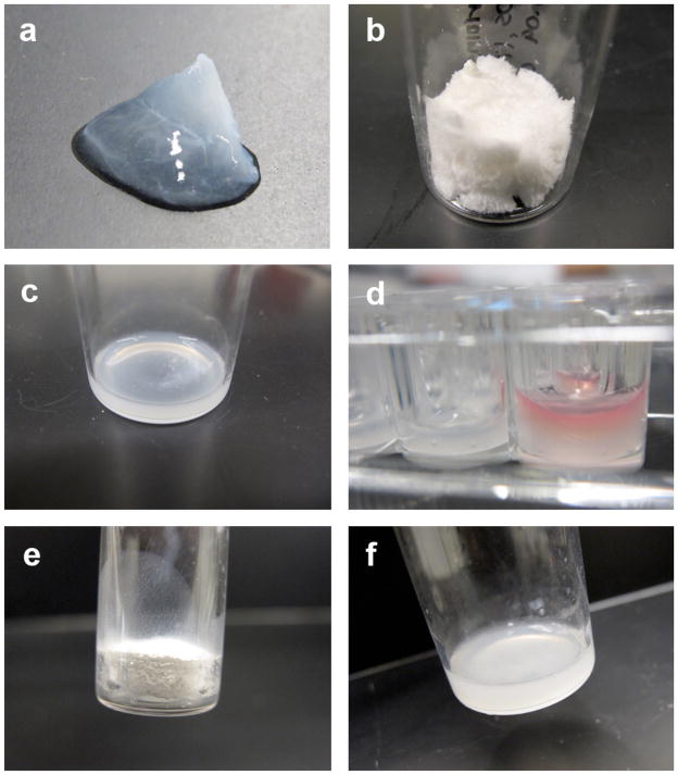
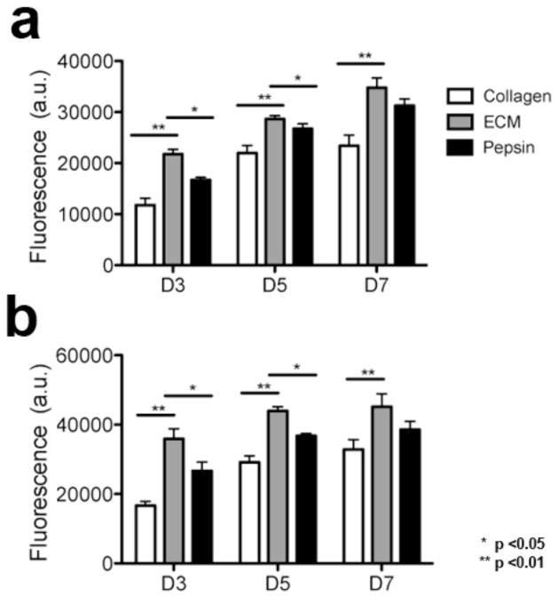
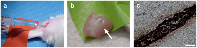
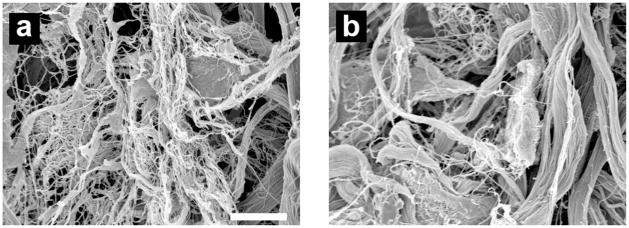
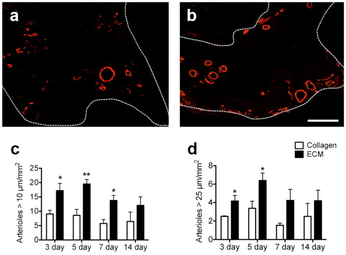
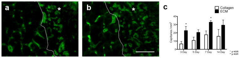
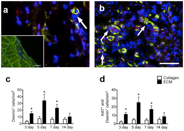
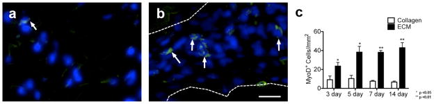
Peripheral artery disease (PAD) currently affects approximately 27 million patients in Europe and North America, and if untreated, may progress to the stage of critical limb ischemia (CLI), which has implications for amputation and potential mortality. Unfortunately, few therapies exist for treating the ischemic skeletal muscle in these conditions. Biomaterials have been used to increase cell transplant survival as well as deliver growth factors to treat limb ischemia; however, existing materials do not mimic the native skeletal muscle microenvironment they are intended to treat. Furthermore, no therapies involving biomaterials alone have been examined. The goal of this study was to develop a clinically relevant injectable hydrogel derived from decellularized skeletal muscle extracellular matrix and examine its potential for treating PAD as a stand-alone therapy by studying the material in a rat hindlimb ischemia model. We tested the mitogenic activity of the scaffold's degradation products using an in vitro assay and measured increased proliferation rates of smooth muscle cells and skeletal myoblasts compared to collagen. In a rat hindlimb ischemia model, the femoral artery was ligated and resected, followed by injection of 150 µL of skeletal muscle matrix or collagen 1 week post-injury. We demonstrate that the skeletal muscle matrix increased arteriole and capillary density, as well as recruited more desmin-positive and MyoD-positive cells compared to collagen. Our results indicate that this tissue-specific injectable hydrogel may be a potential therapy for treating ischemia related to PAD, as well as have potential beneficial effects on restoring muscle mass that is typically lost in CLI.


Figures

Figure 1
Figure 1
Decellularization and tissue processing. (A) Decellularized skeletal muscle matrix. (B) Lyophilized skeletal muscle matrix prior to milling. (C) Digested skeletal muscle matrix. (D) In vitro gel of the skeletal muscle matrix with media on top in right well (E) Skeletal muscle matrix that has been digested and re-lyophilized. (F) Re-lyophilized skeletal muscle matrix resuspended using only sterile water.
Figure 2
Figure 2
In vitro mitogenic activity assay. (A) Rat aortic smooth muscle cells and (B) C2C12 skeletal myoblasts were cultured using growth media with the addition of degraded skeletal muscle matrix, collagen, or pepsin. Proliferation rate was increased for both cell types when cultured in the presence of skeletal muscle matrix degradation products.
Figure 3
Figure 3
Rheological data. A representative trace of the storage (G′) and loss (G″) moduli for the skeletal muscle matrix gel is shown.
Figure 4
Figure 4
Skeletal muscle matrix delivery and gelation in situ. (A) Intramuscular injection of the skeletal muscle matrix material. (B) Gelation of the skeletal muscle matrix in situ after 20 minutes as seen after excision of the muscle; arrow denotes the white matrix (C) DAB staining of the biotin-labeled skeletal muscle matrix that gelled within the muscle. Scale bar at 200 μm.
Figure 5
Figure 5
Scanning electron microscopy. Micrograph of a cross-section of skeletal muscle matrix formed (A) in vitro, and (B) 20 minutes post-subcutaneous injection. Note the formation of the assembled fibers on the nano- and micro- scale. Scale bar at 100 μm.
Figure 6
Figure 6
Quantification of arterioles. (A) Collagen and (B) skeletal muscle matrix injection regions stained with anti-alpha-SMA (red) to determine arteriole formation. Vessels with a clear lumen are seen within the injection region at 5 days. Scale bar at 100 μm. Quantification of the vessel density at 3, 5, 7, and 14 days for vessels with a lumen (C) >10 μm or (D) >25 μm demonstrated that the skeletal muscle matrix increased neovascularization. Vessels were, on average, larger in the skeletal muscle matrix when compared to collagen.
Figure 7
Figure 7
Quantification of endothelial cell recruitment. (A) Collagen and (B) skeletal muscle matrix injection regions stained with isolectin (green) to assess endothelial cell infiltration at 5 days. Scale bar at 100 μm. * and dotted line denote area of material (C) Endothelial cell infiltration at 3, 5, 7, and 14 days was similar across all four time points, but was significantly greater in the skeletal muscle matrix injection region at 3 and 7 days post-injection.
Figure 8
Figure 8
Proliferating muscle cell recruitment. (A) Collagen injection region and (B) skeletal muscle matrix injection region at 5 days with desmin-stained cells (green) co-labeled with Ki67 (red). Arrows denote desmin and Ki67 positive cells. Scale bar at 20 μm. Insert shows positive desmin staining of healthy skeletal muscle, scale bar at 100 μm. (C) Quantification of desmin-positive cells in the skeletal muscle matrix compared to collagen normalized to area. Note that there are significantly more desmin-positive cells in the skeletal muscle matrix. (D) Of these desmin-positive cells, a majority of the cells are proliferating as seen by Ki67 co-labeling.
Figure 9
Figure 9
Muscle progenitor infiltration. MyoD positive cells (green) in (A) collagen and (B) skeletal muscle matrix injection regions at 5 days. Area of injection is denoted by the dotted line. Scale bar at 20 μm. (C) Graph of MyoD-positive cells normalized to the area for the injection region. The number of MyoD-positive cells was significantly higher in the skeletal muscle matrix regions at all time points.

Similar articles

Cited by

References

    1. Alev C, Ii M, Asahara T. Endothelial progenitor cells: a novel tool for the therapy of ischemic diseases. Antioxid Redox Signal. 2011;15:949–965. - PubMed
    1. Bach AD, Arkudas A, Tjiawi J, Polykandriotis E, Kneser U, Horch RE, Beier JP. A new approach to tissue engineering of vascularized skeletal muscle. J Cell Mol Med. 2006;10:716–726. - PMC - PubMed
    1. Badylak SF. The extracellular matrix as a biologic scaffold material. Biomaterials. 2007;28:3587–3593. - PubMed
    1. Badylak SF, Freytes DO, Gilbert TW. Extracellular matrix as a biological scaffold material: Structure and function. Acta Biomater. 2009;5:1–13. - PubMed
    1. Badylak SF, Gilbert TW. Immune response to biologic scaffold materials. Semin Immunol. 2008;20 :109–116. - PMC - PubMed

Publication types