. 2012 Aug 10;287(33):27275-89.
doi: 10.1074/jbc.M112.364125. Epub 2012 Jun 3.

Structural basis for WDR5 interaction (Win) motif recognition in human SET1 family histone methyltransferases

Affiliations

Structural basis for WDR5 interaction (Win) motif recognition in human SET1 family histone methyltransferases

Venkatasubramanian Dharmarajan et al. J Biol Chem. .

Abstract

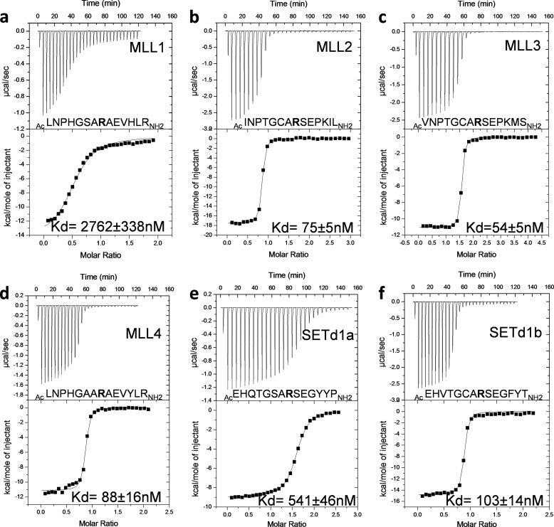
Translocations and amplifications of the mixed lineage leukemia-1 (MLL1) gene are associated with aggressive myeloid and lymphocytic leukemias in humans. MLL1 is a member of the SET1 family of histone H3 lysine 4 (H3K4) methyltransferases, which are required for transcription of genes involved in hematopoiesis and development. MLL1 associates with a subcomplex containing WDR5, RbBP5, Ash2L, and DPY-30 (WRAD), which together form the MLL1 core complex that is required for sequential mono- and dimethylation of H3K4. We previously demonstrated that WDR5 binds the conserved WDR5 interaction (Win) motif of MLL1 in vitro, an interaction that is required for the H3K4 dimethylation activity of the MLL1 core complex. In this investigation, we demonstrate that arginine 3765 of the MLL1 Win motif is required to co-immunoprecipitate WRAD from mammalian cells, suggesting that the WDR5-Win motif interaction is important for the assembly of the MLL1 core complex in vivo. We also demonstrate that peptides that mimic SET1 family Win motif sequences inhibit H3K4 dimethylation by the MLL1 core complex with varying degrees of efficiency. To understand the structural basis for these differences, we determined structures of WDR5 bound to six different naturally occurring Win motif sequences at resolutions ranging from 1.9 to 1.2 Å. Our results reveal that binding energy differences result from interactions between non-conserved residues C-terminal to the Win motif and to a lesser extent from subtle variation of residues within the Win motif. These results highlight a new class of methylation inhibitors that may be useful for the treatment of MLL1-related malignancies.


Figures

FIGURE 1.
FIGURE 1.
Domain architecture of human MLL1. Top, conserved features in MLL1 are indicated as follows: MBM, menin binding motif; LBD, LEDGF binding domain; AT-Hooks and CXXC, DNA binding domains; PHD, plant homeodomain; BD, bromodomain; TAD, Transcriptional Activation Domain; FYRN and FYRC, N-terminal and C-terminal phenylalanine-tyrosine-rich regions, respectively. Bottom, ClustalW2 multiple sequence alignment (66) for the Win motif peptides from human SET1 family members MLL1 (UniProtKB/Swiss-Prot accession number Q03164), MLL2 (UniProtKB/Swiss-Prot accession number O14686), MLL3 (UniProtKB/Swiss-Prot number Q8NEZ4), MLL4 (UniProtKB/Swiss-Prot accession number Q9UMN6), SETd1a (UniProtKB/Swiss-Prot accession number O15047), and SETd1b (UniProtKB/Swiss-Prot number Q9UPS6). BLOSUM matrix (67) was used for the alignment. The conserved Win motif residues are highlighted in bold. Conserved residues are denoted underneath the alignment by an asterisk (*), conservative substitutions are denoted by a colon (:), and semiconservative substitutions are denoted by a period (.). The amino acid sequences of conserved Win motifs are boxed.
FIGURE 2.
FIGURE 2.
Arg-3765 of MLL1 is required for the co-immunoprecipitation (IP) of WDR5, RbBP5, and Ash2L from mammalian cells. In lanes 1–3, Western blots using antibodies against WDR5, RbBP5, Ash2L, and Myc show that equal amounts of the MLL1 wild-type (lane 2) and MLL1 R3765A mutant (lane 3) proteins are present in the input. Lane 1 shows the vector only control. Lanes 4–6 show Western blots from samples immunoprecipitated with the anti-Myc antibody. WDR5, RbBP5, and Ash2L are co-immunoprecipitated along with wild-type MLL1 (lane 5) but are absent in the MLL1 R3765A mutant (lane 6) or vector-only control (lane 4).
FIGURE 3.
FIGURE 3.
ITC data showing that peptides derived from the MLL1 Win motif have similar affinities for WDR5. a–d, ITC data for WDR5 binding to MLL1(3755–3773) (a), MLL1(3758–3771) (b), and MLL1(3762–3773) (c) Win motif peptides. a–c, upper panels, show heat of binding plotted as a function of time. Lower panels show the binding isotherms fit to a one-site binding model. d, peptide sequence and dissociation constants (Kd ± S.E.) derived from the fits are indicated.
FIGURE 4.
FIGURE 4.
ITC data showing that Win motif peptides derived from human SET1 family members have differential affinities for WDR5. a–f, ITC data for WDR5 binding to MLL1 (a), MLL2 (b), MLL3 (c), MLL4 (d), SETd1a (e), and SETd1b (f) Win motif peptides. Upper panels show heat of binding plotted as a function of time. Lower panels show the binding isotherms fit to a one-site binding model. Peptide sequence and dissociation constants (Kd ± S.E.) derived from the fits are indicated.
FIGURE 5.
FIGURE 5.
SET1 family Win motif peptides inhibit the H3K4 dimethylation activity of the MLL1 core complex. a and b, MALDI-TOF assays for the H3K4 methylation activity catalyzed by the MLL1 core complex (MLL3745, WDR5, RbBP5, and Ash2L). MALDI-TOF assays were carried out with 7.3 μm MLL1 core complex for 12 h in the absence or presence of increasing concentrations of MLL1 (green), MLL2 (pink), MLL3 (yellow), MLL4 (orange), SETd1a (cyan), or SETd1b (dark blue) Win motif peptides. a, relative H3K4 dimethylation levels in the presence of increasing concentrations of Win motif peptides or control p53 peptide. Error bars represent the variation observed in two or three independent experiments. The data for the p53 peptide did not converge with the model used for determining IC50; therefore the data were not fitted. b, representative inhibition data for the MLL3 Win motif peptide are shown with concentrations ranging from 0 to a 30-fold molar excess of peptide to that of the MLL1 core complex. Unmod, unmodified; Mono, monomethylated; Di, dimethylated; Tri, trimethylated.
FIGURE 6.
FIGURE 6.
Human SET1 family Win motif peptides bind WDR5 using the same arginine-binding pocket. a–h, the WDR5 molecule is shown as a surface representation (gray) and the different Win motif peptides are shown in different colors. a, superposition of Win motif peptides shown with a schematic representation. b, cut-away view of the arginine-binding pocket of WDR5 is shown. All Win motif peptides insert the conserved arginine into the central tunnel in WDR5. The side chain of the conserved arginine is shown as a stick representation. c–f, simulated annealing FoFc omit maps showing the different Win motif peptides bound to WDR5. MLL1 (c) and MLL3 (e) Win motif peptides are contoured at 2σ, and MLL2 (d), MLL4 (f), SETd1a (g), and SETd1b (h) Win peptides are contoured at 3σ. The peptides are shown as sticks and color-coded as follows: MLL1, green; MLL2, pink; MLL3, yellow; MLL4, orange; SETd1a, cyan; and SETd1b, dark blue).
FIGURE 7.
FIGURE 7.
Conserved Win motif residues adopt structurally similar conformations. Shown is model superposition of the six conserved Win motif residues from different WDR5·Win motif peptide co-crystal structures. Conserved Win motif residues are indicated and numbered with respect to the conserved arginine (position 0). Refer to Table 1 for residue numbering. WDR5 molecule is shown as a surface representation (gray), and the different Win motif peptides are shown as sticks (color-coded as in Fig. 5).
FIGURE 8.
FIGURE 8.
Structural comparison of N-terminal flanking residues from different SET1 family Win motif peptides. a–f, N-terminal flanking residues (−3 and −4) of MLL2 (b), MLL3 (c), MLL4 (d), and SETd1a (e) Win motif peptides adopt a U-shaped conformation where the concave surface interacts with the +2 glutamate side chain. Red dashes show hydrogen bonds between the −4 and +2 residues. Refer to the text for a more detailed description.
FIGURE 9.
FIGURE 9.
Superposition of SET1 family Win motif peptides showing the structural differences at the C-terminal flanking residues. a, +4 amino acid interactions with the A- and B-pockets of WDR5. WDR5 residues that form the A- (red circle) and B (blue circle)-pockets are shown as sticks (white). Different Win peptides are color-coded as in Figs. 6–8. b, representative superposition of A-pocket peptide (MLL1; green) and B-pocket peptide (MLL2; magenta) at position +3. The +4 residue in B-pocket peptides forms an intermolecular hydrogen bond (red dotted line) with the main chain carbonyl of WDR5 residue Lys-259. c, superposition of A- (Phe-149, Asp-172, Pro-173, and Tyr-191) and B-pocket (Tyr-191, Pro-216, and Leu-234) WDR5 residues is shown for all structures without Win peptides. The largest differences are observed in the side chains of Tyr-191 and Lys-259. The WDR5 residues are color-coded as follows: MLL1, green; MLL2, pink; MLL3, yellow; MLL4, orange; SETd1a, cyan; and SETd1b, dark blue.
FIGURE 10.
FIGURE 10.
Structural differences between MLL1 and MLL4 Win motif peptides contribute to the differences in binding affinity to WDR5. a and b, superposition of MLL1 and MLL4 Win motif peptides. a, view of −2 position of MLL1 (green) and MLL4 (orange) Win motif peptides. WDR5 residues are shown as line representations. b, view of the +4 amino acid in MLL1 (green) and MLL4 (orange) structures. MLL4 residue Tyr-2515 (+4 position) sits in the A-pocket and forms a direct hydrogen bond (pink dotted line) with the WDR5 residue Asp-172. c, IC50 plots for the inhibition of the H3K4 dimethylation activity of the fully assembled MLL1 core complex by the wild-type and mutant MLL1 Win motif peptides. For comparison, MLL4 and the control p53 peptide plots are also shown. The methylation reactions were carried out as described in Fig. 5. Data presented are an average of two independent experiments and the error bars represent standard deviation from the mean. d, IC50, Ki, and the standard free energy of binding values derived from the fits to the data in c are summarized. ΔΔG0′ values represent the difference in free energy of binding between wild-type and mutant MLL1 Win motif peptides.

Similar articles

Cited by

References

    1. Dillon S. C., Zhang X., Trievel R. C., Cheng X. (2005) The SET-domain protein superfamily: protein lysine methyltransferases. Genome Biol. 6, 227. - PMC - PubMed
    1. Cosgrove M. S. (2006) PHinDing a new histone “effector” domain. Structure 14, 1096–1098 - PubMed
    1. Flanagan J. F., Mi L. Z., Chruszcz M., Cymborowski M., Clines K. L., Kim Y., Minor W., Rastinejad F., Khorasanizadeh S. (2005) Double chromodomains cooperate to recognize the methylated histone H3 tail. Nature 438, 1181–1185 - PubMed
    1. Pray-Grant M. G., Daniel J. A., Schieltz D., Yates J. R., 3rd, Grant P. A. (2005) Chd1 chromodomain links histone H3 methylation with SAGA- and SLIK-dependent acetylation. Nature 433, 434–438 - PubMed
    1. Santos-Rosa H., Schneider R., Bernstein B. E., Karabetsou N., Morillon A., Weise C., Schreiber S. L., Mellor J., Kouzarides T. (2003) Methylation of histone H3 K4 mediates association of the Isw1p ATPase with chromatin. Mol. Cell 12, 1325–1332 - PubMed

Publication types

MeSH terms