. 2012 Aug;32(15):3153-63.
doi: 10.1128/MCB.00161-12. Epub 2012 Jun 4.

Ubiquitylation-dependent negative regulation of WASp is essential for actin cytoskeleton dynamics

Affiliations

Ubiquitylation-dependent negative regulation of WASp is essential for actin cytoskeleton dynamics

Barak Reicher et al. Mol Cell Biol. 2012 Aug.

Abstract

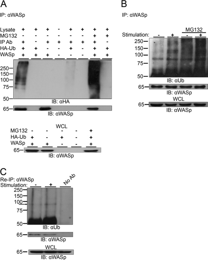
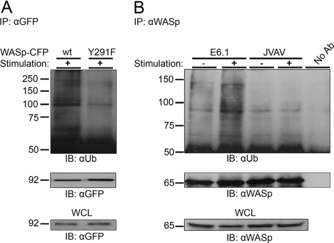
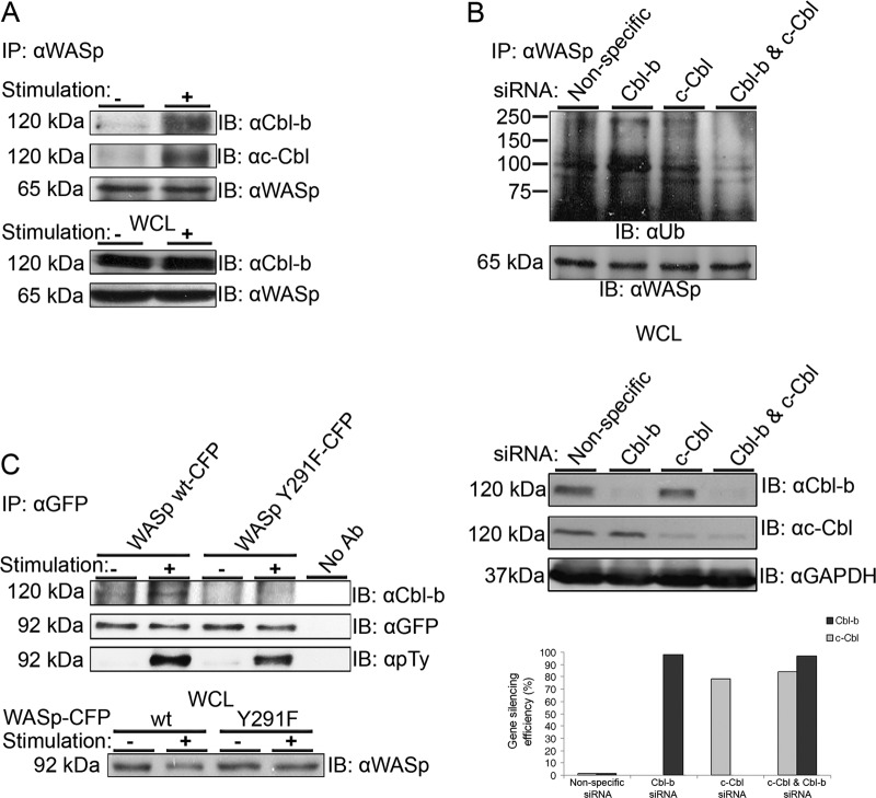
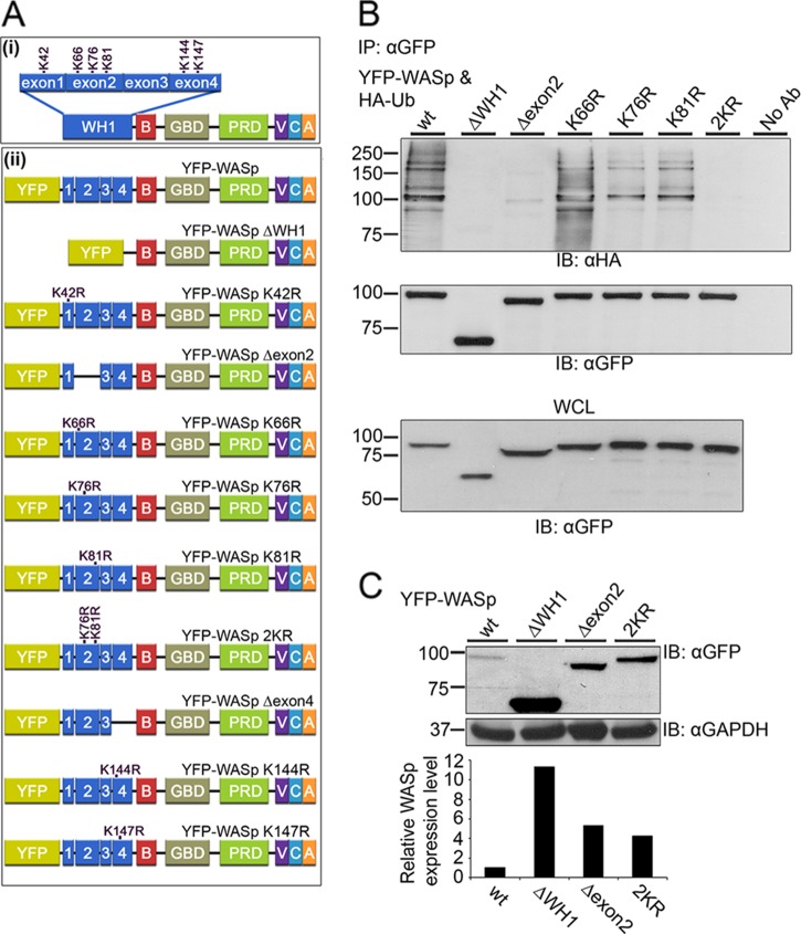
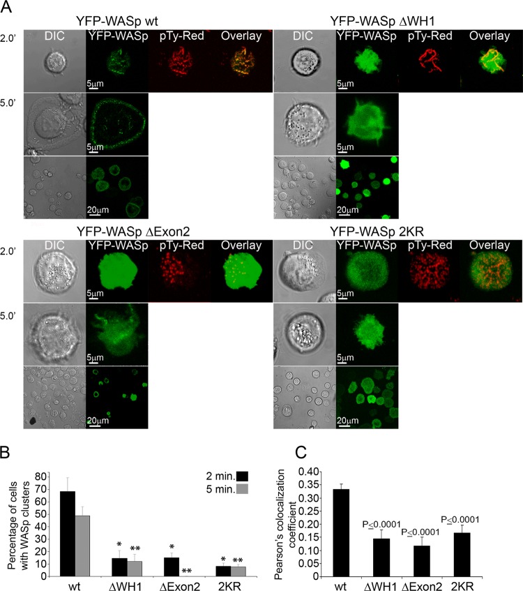
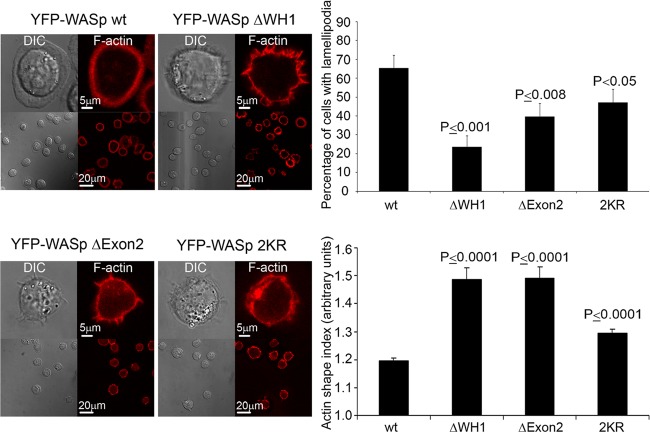
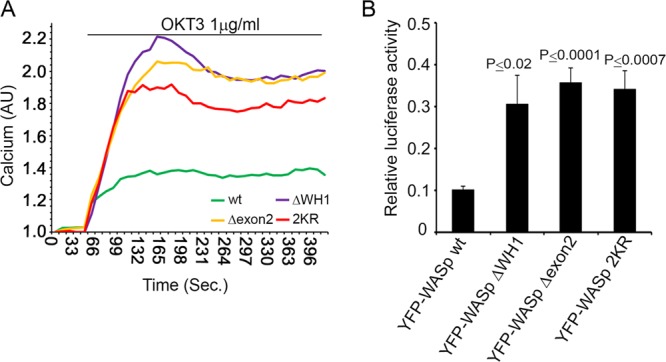
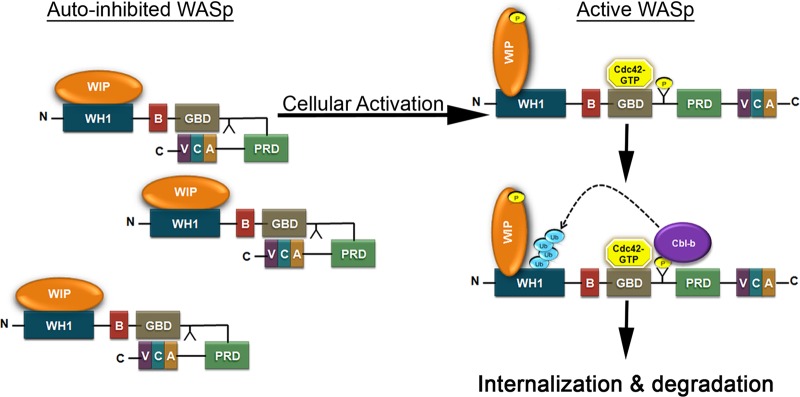
The Wiskott-Aldrich syndrome protein (WASp) is a key regulator of actin dynamics during cell motility and adhesion, and mutations in its gene are responsible for Wiskott-Aldrich syndrome (WAS). Here, we demonstrate that WASp is ubiquitylated following T-cell antigen receptor (TCR) activation. WASp phosphorylation at tyrosine 291 results in recruitment of the E3 ligase Cbl-b, which, together with c-Cbl, carries out WASp ubiquitylation. Lysine residues 76 and 81, located at the WASp WH1 domain, which contains the vast majority of WASp gene mutations, serve as the ubiquitylation sites. Disruption of WASp ubiquitylation causes WASp accumulation and alters actin dynamics and the formation of actin-dependent structures. Our data suggest that regulated degradation of activated WASp might be an efficient strategy by which the duration and localization of actin rearrangement and the intensity of T-cell activation are controlled.


Figures

Fig 1
Fig 1
Ubiquitylation of WASp. (A) WASp was immunoprecipitated (IP: αWASp) from 293T cells transiently expressing WASp and HA-tagged ubiquitin (HA-Ub), WASp alone, or HA-Ub alone. Cells coexpressing WASp and HA-Ub were left untreated or incubated in the presence of MG132. The immunoprecipitates were resolved by SDS-PAGE and immunoblotted for ubiquitin using the anti-HA antibody (IB: αHA) and for WASp using anti-WASp antibody. Ubiquitylated WASp bands appear as a smear of bands above the molecular mass of ∼81 kDa. Whole-cell lysates (WCL) of the indicated samples were also blotted for WASp (lower panel). (B) Jurkat T cells were left unstimulated (−) or stimulated with anti-CD3 antibody (+). MG132-treated cells were also left unstimulated (−) or stimulated (+). Cell lysates were immunoprecipitated using anti-WASp antibody and immunoblotted for ubiquitin. Ubiquitylated endogenous WASp bands appear as a smear of bands around the molecular mass of ∼81 kDa. WCL of the indicated samples were also blotted for WASp (lower panel). Data shown are representative of five independent experiments. (C) Jurkat T cells were left unstimulated (−) or stimulated with anti-CD3 antibody (+). Cell lysates were immunoprecipitated using anti-WASp antibody, followed by boiling in a denaturing buffer to separate WASp-associated proteins. After dilution in cell lysis buffer, WASp was reimmunoprecipitated (Re-IP: αWASp). The precipitates from the denatured/renatured samples were immunoblotted for ubiquitin and WASp (IB: αUb and IB: αWASp, respectively). Ubiquitylated endogenous WASp bands are indicated with an arrow. WCL of the indicated samples were also blotted for WASp (lower panel). Data shown are representative of two independent experiments.
Fig 2
Fig 2
TCR-induced WASp phosphorylation on the tyrosine 291 site is required for WASp ubiquitylation. (A) Jurkat T cells transiently expressing WASp wt-CFP or the Y291F-CFP mutant form of WASp were stimulated (+), and WASp-CFP was immunoprecipitated using anti-GFP antibody. Immunoblotting analysis for ubiquitin and for WASp-CFP with antiubiquitin and anti-GFP antibodies, respectively, was performed. Ubiquitylated WASp-CFP bands appear as a smear of bands around the molecular mass of ∼100 kDa. WCL of the indicated samples were also blotted for WASp-CFP (lower panel). Data shown are representative of three independent experiments. (B) VAV1, a guanine nucleotide exchange factor, is required for WASp ubiquitylation. WASp ubiquitylation was examined in Jurkat E6.1 and in VAV1-deficient T cells (JVAV). WASp was immunoprecipitated with anti-WASp antibody from unstimulated or TCR-stimulated cell lysates. WASp precipitates were analyzed by immunoblotting with anti-Ub antibody. Precipitates were also analyzed by WASp immunoblotting. Ubiquitylated endogenous WASp bands appear as a smear of bands around the molecular mass of ∼81 kDa. WCL of the indicated samples were also blotted for WASp (lower panel). Data shown are representative of three independent experiments.
Fig 3
Fig 3
The E3 ligases, Cbl-b and c-Cbl, physically associate with endogenous WASp in T cells. (A) E6.1 T cells were left unstimulated or were stimulated using anti-TCR antibody (C3O5). Cells were lysed, followed by immunoprecipitation of WASp (IP: αWASp). The immunoprecipitates and the WCL were resolved on SDS-PAGE and immunoblotted for Cbl-b (upper panel), c-Cbl (middle panel), and WASp (lower panel). Data shown are representative of six independent experiments. (B) Gene silencing of both Cbl-b and c-Cbl reduces WASp ubiquitylation. E6.1 T cells were transfected with siRNA specific to human Cbl-b, c-Cbl, or a mixture of both as well as with a nontargeting siRNA control (nonspecific). After 48 h, cell lysates were prepared and analyzed for Cbl protein levels. The gene-silencing efficiencies were calculated by ImageJ densitometry results for Cbl-b and c-Cbl after normalization against the GAPDH values and comparison to the values of the negative controls (lower panel). Endogenous WASp was immunoprecipitated by anti-WASp, and the membranes were blotted with anti-Ub and anti-WASp. Ubiquitylated endogenous WASp bands appear as a smear of bands around the molecular mass of ∼81 kDa. Data shown are representative of six independent experiments. (C) Phosphorylation of WASp on the tyrosine 291 site is required for its interaction with Cbl-b. E6.1 T cells stably expressing WASp (wt-CFP) or a mutant form of WASp (Y291F-CFP) were stimulated (+) and immunoprecipitated for WASp-CFP using anti-GFP antibody. Immunoblotting with anti-Cbl-b, -GFP, and -phosphotyrosine (pTy) was performed. WCL of the indicated samples were also blotted for WASp (lower panel). Data shown are representative of three independent experiments.
Fig 4
Fig 4
Identification of WASp domains and lysine residues required for its ubiquitylation. (Ai) Schematic illustration of WASp structural domains. WH1, WASp homology 1 domain; B, basic region; GBD, GTPase-binding domain; PRD, proline-rich domain; VCA, verprolin homology/central region/acidic region. The WASp WH1 domain consists of four exons containing six lysines. (Aii) Schemes of WASp wt and WASp mutant forms: WH1-deleted WASp (ΔWH1), exon 2-deleted WASp (Δexon2), exon 4-deleted WASp (Δexon4), and point-mutated WASp with the indicated lysine residues replaced by arginine. (B) 293T cells were cotransfected with constructs encoding HA-tagged ubiquitin together with YFP-WASp wt or with YFP-WASp mutant forms described above (Aii). YFP-WASp was immunoprecipitated using anti-GFP antibody; complexes were resolved by SDS-PAGE and immunoblotted for ubiquitin using the anti-HA and anti-GFP antibodies. Ubiquitylated YFP-WASp bands appear as a smear of bands around the molecular mass of ∼100 kDa. WCL of the indicated samples were also blotted for YFP-WASp (lower panel). Data shown are representative of five independent experiments. (C) Accumulation of WASp ΔWH1, WASp Δexon2, and WASp 2KR mutant forms. Jurkat T cells expressing YFP-WASp wt or the YFP-WASp mutant forms ΔWH1, Δexon2, and 2KR were sorted for GFP-positive cells. Anti-CD3 antibody-stimulated cells were lysed and analyzed for WASp protein levels by immunoblotting with anti-GFP antibody or anti-GAPDH as a loading control. Densitometric analysis of the bands presented was performed by using ImageJ and normalized by the GAPDH densitometry values. Data shown are representative of five independent experiments.
Fig 5
Fig 5
Accumulation of WASp ΔWH1, WASp Δexon2, and WASp 2KR mutant forms causes aberrant WASp cluster formation and T-cell spreading. (A) The distribution of WASp wt was compared to those of the YFP-WASp mutant forms ΔWH1, Δexon2, and 2KR in activated T cells. E6.1 T cells expressing YFP-WASp wt or mutants (green) were plated on a stimulatory coverslip and then fixed and stained with antiphosphotyrosine (red). Right panels display collected images at 2 and 5 min into the spreading process. The images of YFP-WASp wt were taken under the same conditions as those of the YFP-WASp ΔWH1, WASp Δexon2, and WASp 2KR proteins. Left panels represent the DIC channel. Images on the bottom show the entire field of cells. (B) The percentage of cells that produced WASp clusters in cells expressing YFP-WASp wt was compared to that in cells expressing the WASp mutant forms at 2 and 5 min into the spreading process. A t test analysis between WASp wt and the WASp mutants was performed at 2 (*) and 5 (**) min (P < 0.0001 for both time points). (C) Colocalization analysis based on Pearson's colocalization coefficients. Image analysis was performed on more than 50 cells for each experimental group. Data shown are representative of three independent experiments.
Fig 6
Fig 6
WASp ΔWH1-, WASp Δexon2-, or WASp 2KR-expressing cells form mainly membrane spike structures and fewer lamellipodia. E6.1 T cells expressing YFP-WASp wt, WASp ΔWH1, WASp Δexon2, or WASp 2KR were sorted for YFP-positive cells, plated, fixed after 5 min of activation, and stained with phalloidin. Cells producing lamellipodia were counted, and their percentage of the total cells was calculated. Actin shape index was analyzed by IPLab software as described in Materials and Methods. The images of at least 50 cells (for each cell type) were analyzed. A t test analysis between WASp wt and the WASp mutants was performed with a P value of <0.05, as indicated within the figure.
Fig 7
Fig 7
Abrogation of WASp negative regulation enhances T-cell activation. (A) Jurkat T cells were transfected with YFP-WASp wt or mutant forms. YFP-WASp-positive cells were sorted and loaded with 5 μg calcium-sensitive Indo-1-AM as described in Materials and Methods. Cells were then analyzed for intracellular calcium levels by spectrofluorometry following stimulation with anti-CD3 (OKT3, 1 μg/ml) antibody. Data shown are representative of four independent experiments. (B) Jurkat T cells were cotransfected with YFP-WASp wt or its mutant forms, ΔWH1, Δexon2, or 2KR, and the NFAT luciferase reporter plasmid. Cells were lysed and assayed for luciferase activity. NFAT luciferase activity obtained upon anti-TCR stimulation (2 μg/ml) was normalized for transfection efficiency using values from the Renilla-luciferin reaction and expressed as a fraction of the maximal stimulation (PMA and ionomycin). A t test analysis between WASp wt and the WASp mutants is presented. Data shown are representative of at least five independent experiments.
Fig 8
Fig 8
Schematic representation of the molecular mechanism of WASp ubiquitylation. In naive T cells, WASp tightly associates with WIP via its N terminus and exon 2-encoded fragment (aa 56 to 102). WIP masks lysine residues 76 and 81 and inhibits their modification via ubiquitin, and thus, WASp accumulates within the T cell. Upon cellular activation, the Rho family GEF VAV1 activates Cdc42, which binds to the WASp GBD domain, thereby releasing WASp from its autoinhibitory conformation and exposing the VCA domain. The VCA domain of WASp binds to the Arp2/3 complex, promoting local actin polymerization. Phosphorylation of WASp on Y291 by PTK increases the sensitivity of WASp to Cdc42. WASp-WIP interactions are altered following activation. Phosphorylation of WASp on Y291 mediates its interaction with the E3 ligases. This event leads to the ubiquitylation of WASp on lysine residues 76 and 81 and to its degradation.

References

    1. Badour K, et al. 2004. Fyn and PTP-PEST-mediated regulation of Wiskott-Aldrich syndrome protein (WASp) tyrosine phosphorylation is required for coupling T cell antigen receptor engagement to WASp effector function and T cell activation. J. Exp. Med. 199:99–112 - PMC - PubMed
    1. Badour K, Zhang J, Siminovitch KA. 2004. Involvement of the Wiskott-Aldrich syndrome protein and other actin regulatory adaptors in T cell activation. Semin. Immunol. 16:395–407 - PubMed
    1. Badour K, Zhang J, Siminovitch KA. 2003. The Wiskott-Aldrich syndrome protein: forging the link between actin and cell activation. Immunol. Rev. 192:98–112 - PubMed
    1. Balagopalan L, et al. 2007. c-Cbl-mediated regulation of LAT-nucleated signaling complexes. Mol. Cell Biol. 27:8622–8636 - PMC - PubMed
    1. Barda-Saad M, et al. 2005. Dynamic molecular interactions linking the T cell antigen receptor to the actin cytoskeleton. Nat. Immunol. 6:80–89 - PubMed

Publication types

MeSH terms

LinkOut - more resources