. 2012 Aug;32(15):3176-86.
doi: 10.1128/MCB.00086-12. Epub 2012 Jun 4.

The Abl and Arg kinases mediate distinct modes of phagocytosis and are required for maximal Leishmania infection

Affiliations

The Abl and Arg kinases mediate distinct modes of phagocytosis and are required for maximal Leishmania infection

Dawn M Wetzel et al. Mol Cell Biol. 2012 Aug.

Abstract

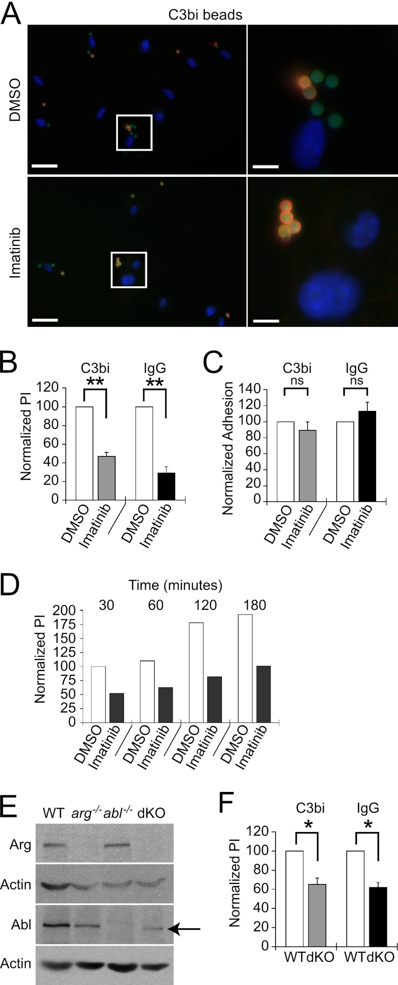
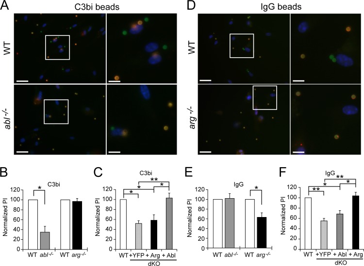
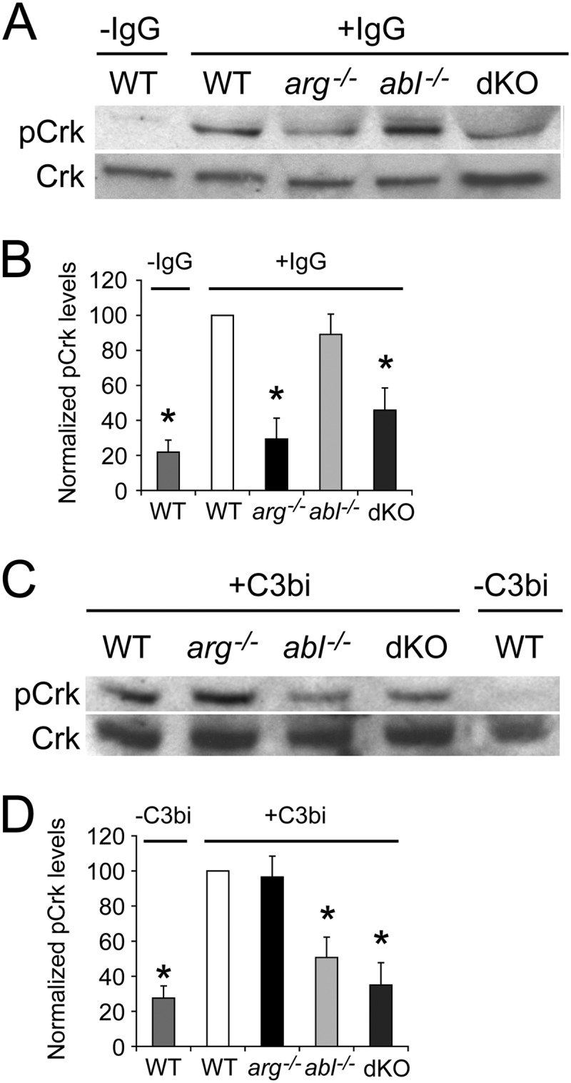
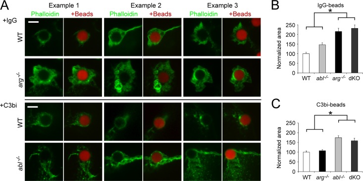
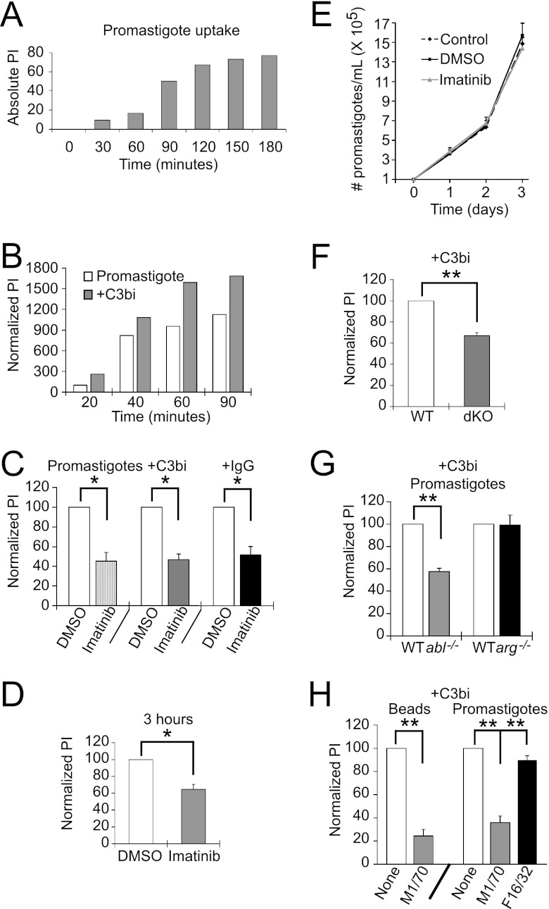
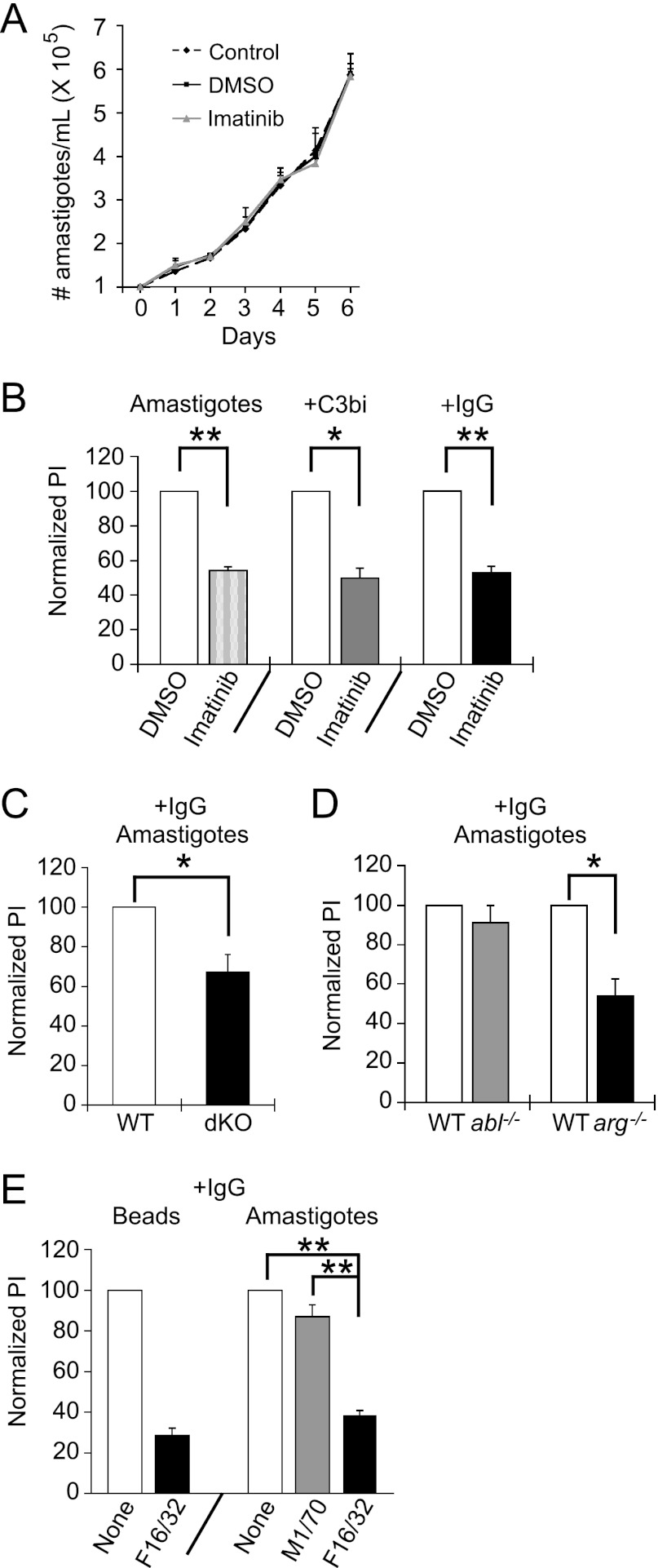
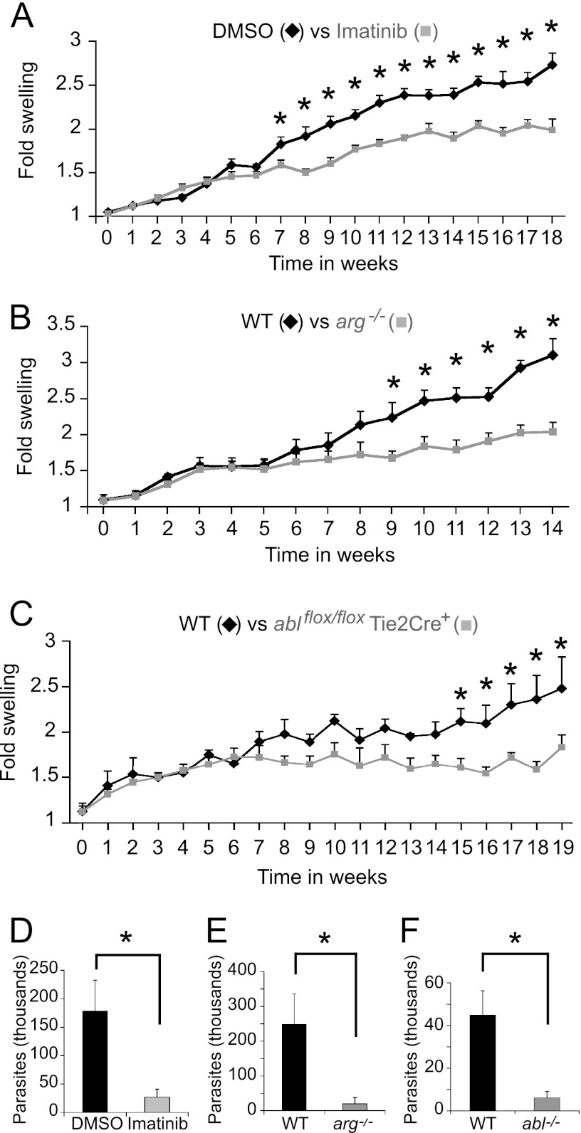
Leishmania, an obligate intracellular parasite, binds several receptors to trigger engulfment by phagocytes, leading to cutaneous or visceral disease. These receptors include complement receptor 3 (CR3), used by promastigotes, and the Fc receptor (FcR), used by amastigotes. The mechanisms mediating uptake are not well understood. Here we show that Abl family kinases mediate both phagocytosis and the uptake of Leishmania amazonensis by macrophages (Ms). Imatinib, an Abl/Arg kinase inhibitor, decreases opsonized polystyrene bead phagocytosis and Leishmania uptake. Interestingly, phagocytosis of IgG-coated beads is decreased in Arg-deficient Ms, while that of C3bi-coated beads is unaffected. Conversely, uptake of C3bi-coated beads is decreased in Abl-deficient Ms, but that of IgG-coated beads is unaffected. Consistent with these results, Abl-deficient Ms are inefficient at C3bi-opsonized promastigote uptake, and Arg-deficient Ms are defective in IgG1-opsonized amastigote uptake. Finally, genetic loss of Abl or Arg reduces infection severity in murine cutaneous leishmaniasis, and imatinib treatment results in smaller lesions with fewer parasites than in controls. Our studies are the first to demonstrate that efficient phagocytosis and maximal Leishmania infection require Abl family kinases. These results highlight Abl family kinase-mediated signaling pathways as potential therapeutic targets for leishmaniasis.


Figures

Fig 1
Fig 1
Abl family kinases are required for optimal phagocytosis. (A and B) Imatinib decreases C3bi- and IgG-opsonized bead uptake. BM Mϕs or RAW 264.7 cells were treated with 3.3 μM imatinib or DMSO for 2 h. Ten C3bi- or IgG-opsonized beads were incubated per Mϕ for 30 min at 37°C. Two-color IF distinguished between intracellular (green) and extracellular (orange) beads. Nuclei are labeled with DAPI. (A) Image of C3bi-opsonized bead uptake by BM Mϕs treated with DMSO (top) or imatinib (bottom). The left panels show representative fields; the right panels show enlarged areas (boxed). Left scale bar, 20 μm; right scale bar, 5 μm. (B) Imatinib inhibits phagocytosis. Bars show the mean phagocytic index (PI) ± standard error (SE) for RAW 264.7 cells treated with 3.3 μM imatinib normalized to the DMSO-treated PI (100%) for each experiment. **, P = 0.0063for C3bi-coated bead uptake by DMSO versus imatinib-treated cells, and P = 0.0093 for IgG-coated bead uptake by DMSO versus imatinib-treated cells, by one-sample t test (n = 3 experiments). (C) Imatinib does not affect adhesion. Bars show percentages of adhered beads per 100 imatinib-treated RAW 264.7 cells compared to DMSO-treated cells from the experiment shown in panel B. (D) The relative decrease in PI in imatinib-treated Mϕs does not change even after 3 h of incubation. Mϕs were treated with DMSO or 3.3 μM imatinib prior to incubation with zymosan particles for the time indicated. The PI is shown for each time point of imatinib-treated Mϕs, normalized to DMSO-treated Mϕs at the first time point. Shown is one representative experiment of two experiments. (E) Immunoblots of Arg and Abl expression in BM Mϕs isolated from WT, arg−/−, abl−/−, or arg−/− ablflox/flox/Tie2-Cre+ (dKO) mice. dKO Mϕs express low levels of a truncated, kinase-inactive form of Abl (indicated by arrow) that migrates 10 kDa faster than full-length Abl on immunoblots. Actin is a loading control. (F) Mϕs lacking Arg with decreased Abl levels (dKO) have phagocytic defects. The graph shows the normalized mean PI ± SE for dKO Mϕs compared to WTLM Mϕs (abbreviated “WT”). *, P = 0.035 for C3bi-coated bead uptake by WTLM versus dKO Mϕs, and P = 0.019 for IgG-coated bead uptake by WTLM versus dKO Mϕs, by one-sample t test.(n = 3 experiments)
Fig 2
Fig 2
Abl facilitates complement-mediated phagocytosis, while Arg governs immunoglobulin-mediated phagocytosis. (A and B) abl−/− Mϕs exhibit defects in complement-mediated phagocytosis. (A) Images of C3bi-opsonized bead uptake by WTLM (top, labeled “WT”) and abl−/− (bottom) Mϕs. Left panels, wide-field images; right panels, enlarged areas (boxed). Mϕs were incubated with beads and processed as in Fig. 1E. Scale bar, 20 μm in the left panels and 7 μm in the right panels. (B) Graph showing the mean PI ± SE for C3bi-opsonized beads by abl−/− and arg−/− Mϕs, normalized to WTLM (100%). *, P = 0.032 for WTLM versus abl−/− Mϕs by one-sample t test (n = 3 experiments). (C) Abl-YFP expression corrects C3bi-mediated phagocytic defects of dKO Mϕs. Shown is the mean PI ± SE for C3bi-opsonized bead uptake by Abl-YFP, Arg-YFP, and YFP-complemented Mϕs, normalized to WTLM Mϕs. *, P < 0.05, and **, P < 0.01, by one-way ANOVA (n = 3 experiments). (D and E) arg−/− Mϕs have defects in immunoglobulin-mediated phagocytosis. (D) Image of IgG-opsonized bead uptake by WTLM (top) and arg−/− (bottom) Mϕs. Coverslip processing, panels, and scale bars are as in panel A. (E) Graph showing mean PI ± SE for IgG-opsonized bead uptake by abl−/− and arg−/− Mϕs, normalized to WTLM Mϕs. *, P = 0.028 for WTLM versus arg−/− Mϕs by one-sample t test (n = 4 experiments). (F) Arg-YFP expression corrects IgG phagocytic defects in dKO Mϕs. Shown is the mean PI ± SE for IgG-coated bead uptake by Abl-YFP-, Arg-YFP-, and YFP-complemented Mϕs, normalized to WTLM Mϕs. *, P < 0.05, and **, P < 0.01 by one-way ANOVA (n = 3 experiments).
Fig 3
Fig 3
Arg and Abl stimulate Crk phosphorylation following engagement of the FcR and CR3 receptors, respectively. (A and B) Phosphorylation of the Abl/Arg substrate CrkII (pCrk) induced upon FcR engagement is decreased in arg−/− and dKO Mϕs compared to WT or abl−/− Mϕs. M-CSF-starved-Mϕs were adhered to uncoated plates (−IgG) or plates coated with mouse IgG1 (+IgG) for 15 min before lysis and processing for immunoblotting with an antibody to pCrk. (A) Representative immunoblot of pCrk (top) and total Crk (bottom) in WT Mϕs (± IgG) and IgG-stimulated arg−/−, abl−/− and dKO Mϕs. (B) Graph showing relative levels of pCrk, normalized to Crk levels, among WT (±IgG), arg−/−, abl−/−, and dKO Mϕs. Relative levels of pCrk for each category have been normalized to the level in IgG-treated WT Mϕs. *, P < 0.05 by ANOVA when pCrk levels in starred categories are compared to IgG-stimulated WT or abl−/− Mϕs (n = 5 experiments). (C and D) CR3 engagement-induced Crk phosphorylation is decreased in abl−/− and dKO Mϕs compared to WT or arg−/− Mϕs. Mϕs were added to uncoated plates (−C3bi) or C3bi-coated plates (+C3bi) for 15 min before processing as described above. (C) Representative immunoblot of pCrk (top) and Crk (bottom) in C3bi-stimulated WT, arg−/−, abl−/−, and dKO Mϕs and unstimulated WT Mϕs. (D) Graph showing relative levels of pCrk, normalized to Crk, among unstimulated WT Mϕs and C3bi-stimulated WT, arg−/−, abl−/−, and dKO Mϕs. *, P < 0.05 by ANOVA if pCrk levels in starred categories are compared to levels in either WT or arg−/− Mϕs (n = 5 experiments).
Fig 4
Fig 4
Loss of Arg or Abl yields large F-actin-rich phagocytic cups during immunoglobulin-mediated or complement-mediated phagocytosis. (A) Three examples of WT and arg−/− Mϕs undergoing immunoglobulin-mediated phagocytosis (+IgG), as well as WT and abl−/− Mϕs undergoing complement-mediated phagocytosis (+C3bi). Mϕs were incubated with red beads and fixed/permeabilized. F-actin was labeled with green phalloidin. Left panel, actin alone; right panel, merged actin and bead. Scale bar, 5 μm. (B) Phagocytic cups in cells undergoing IgG-mediated phagocytosis are larger in arg−/− and dKO Mϕs than in controls. The graph shows the average area of phagocytic cups (normalized to WT) in WT, abl−/−, arg−/−, and dKO Mϕs. *, P < 0.05 for arg−/− or dKO Mϕs compared to WT or abl−/− Mϕs by one-way ANOVA. The difference between WT and abl−/− Mϕs is not statistically significant. (C) Phagocytic cups in cells undergoing complement-mediated phagocytosis are larger in abl−/− and dKO Mϕs than in controls. Normalization performed as for panel B. *, P < 0.05 for abl−/− or dKO Mϕs compared to WT and arg−/− Mϕs by one-way ANOVA.
Fig 5
Fig 5
Abl mediates promastigote engulfment. (A) Time course for cultured promastigote uptake. Promastigotes were incubated with primary Mϕs for the time indicated, and the PI was calculated. Shown is the absolute PI for a representative experiment of two experiments. (B) The effect of C3bi opsonization on uptake decreases over time. Shown is the PI ± SE of cultured and opsonized promastigotes over time, normalized to the PI of cultured promastigotes at 20 min, from a representative experiment of 2. (C) Imatinib decreases uptake of L. amazonensis promastigotes. Mϕs were treated with 3.3 μM imatinib or DMSO. Promastigotes were grown in culture (Promastigotes) or preincubated with mouse serum (+C3bi) or mouse IgG1 anti-gp46 (+IgG) and incubated with Mϕs for 20 min (+C3bi, +IgG) or 90 min (Promastigotes). An antibody to gp46 differentiated internalized (green) from external (orange) promastigotes. The graph shows the average PI ± SE for imatinib-treated Mϕs for cultured promastigotes, C3bi-opsonized promastigotes, and IgG-opsonized promastigotes, each normalized to their respective DMSO-treated Mϕs for 3 experiments. *, P = 0.024 for uptake by DMSO- versus imatinib-treated Mϕs for cultured promastigotes, P = 0.011 for C3bi-opsonized promastigotes, and P = 0.032 for IgG-opsonized promastigotes by one-sample t test. (D) Imatinib continues to decrease parasite uptake even after 3 h. The graph shows the average PI ± SE for imatinib-treated Mϕs, normalized to their respective DMSO-treated Mϕs. *, P < 0.05 by one-tailed t test (n = 3 experiments). (E) Imatinib is not toxic to promastigotes. Shown is a representative experiment of two experiments following promastigotes per ml of triplicate culture in control, DMSO-treated, or 3.3 μM imatinib-treated media over 3 days. (F) C3bi-opsonized promastigote uptake is decreased in Mϕs lacking Abl and Arg. WTLM versus dKO Mϕs were incubated with C3bi-opsonized promastigotes for 20 min. The graph shows the mean PI ± SE by dKO Mϕs, normalized to WTLM Mϕs (labeled WT). **, P = 0.0068 for WTLM versus dKO Mϕs by one-sample t test. (G) Abl mediates C3bi-opsonized promastigote uptake. The graph shows the mean PI ± SE by abl−/− and arg−/− Mϕs, normalized to WTLM Mϕs (labeled WT). **, P = 0.0046 for WTLM versus abl−/− Mϕs by one-sample t test (n = 3 experiments). (H) A CR3-blocking antibody (M1/70) decreases promastigote uptake. RAW 264.7 cells were preincubated with M1/70 or F16/32 (an FcR blocking antibody; both rat IgG2) prior to incubation with C3bi-opsonized promastigotes. The graph shows the mean PI + SE for untreated, M1/70-treated, or F16/32-treated RAW 264.7 cells. **, P < 0.01 for M1/70-treated cells by one-way ANOVA (n = 3 experiments).
Fig 6
Fig 6
Arg mediates amastigote engulfment. (A) Imatinib is not toxic to amastigotes. Shown is a representative experiment of two experiments following the number of amastigotes per ml in control, DMSO-treated, or imatinib-treated medium over 6 days. (B) Imatinib decreases amastigote uptake. Mϕs were pretreated with 3.3 μM imatinib or DMSO. Mouse anti-P8 IgG1 or freshly isolated mouse serum was used to opsonize amastigote surfaces with IgG1 or C3bi. Amastigotes were incubated with Mϕs for 90 min (unopsonized) or 20 min (opsonized). Two-color IF with anti-P8 distinguished internalized from external amastigotes. The graph shows the mean PI ± SE for imatinib-treated Mϕs, normalized to DMSO-treated Mϕs, of opsonized (+ C3bi, + IgG) and unopsonized (Amastigotes) L. amazonensis parasites. P = 0.0024 (**) for DMSO- versus imatinib-treated Mϕs for unopsonized amastigotes, P = 0.013 (*) for C3bi-opsonized amastigotes, and P = 0.0063 (**) for IgG-opsonized amastigotes by one-sample t test. (C) IgG1-opsonized amastigote uptake decreases in Mϕs lacking Abl and Arg. WTLM versus dKO Mϕs were incubated with IgG1-opsonized amastigotes for 20 min. The graph shows the mean PI ± SE by dKO Mϕs, normalized to WTLM Mϕs (labeled WT). *, P = 0.038 for WTLM versus dKO Mϕs by one-sample t test. (D) Arg mediates IgG-opsonized amastigote uptake. Bars show the mean PI ± SE of amastigotes by abl−/− and arg−/− Mϕs, normalized to WTLM (labeled “WT”). *, P = 0.034 for WTLM versus arg−/− Mϕs by one-sample t test. (E) An FcRγIII-blocking antibody (F16/32) decreases amastigote uptake. RAW 264.7 cells were preincubated with F16/32 or M1/70 prior to incubation with IgG1-opsonized amastigotes. The graph shows the mean PI + SE for untreated, M1/70-treated, or F16/32-treated RAW 264.7 cells. **, P < 0.01 for F16/32-treated cells by one-way ANOVA (n = 3 experiments).
Fig 7
Fig 7
Abl family kinases permit efficient infection in a mouse model of cutaneous leishmaniasis. (A) Imatinib-treated mice have smaller lesions than untreated mice. Four to eight C57BL/6 mice per experiment were injected with 1 × 10⁶ L. amazonensis promastigotes in the right hind foot and treated with 200 mg/kg/day of imatinib or DMSO in their drinking water, starting 4 days before infection and continuing until sacrifice. Three separate experiments were performed; shown is a representative experiment containing 8 mice per group. Bars represent the increase in foot size compared to the uninfected foot (normalized to 1) ± SE. *, P < 0.05 by ANOVA. (B) arg−/− mice and (C) ablflox/flox/Tie2-Cre+ mice develop smaller lesions than WTLM when infected with 1 × 10⁶ L. amazonensis promastigotes. Shown in panel B is a cohort of 5 mice per group in a representative experiment performed in duplicate using 4 to 5 mice per group. Shown in panel C is a cohort of 4 mice per group in a representative, duplicated experiment with 4 to 8 mice in each group. *, P < 0.05 by ANOVA. (D) Lesions in imatinib-treated mice contain fewer parasites than lesions in DMSO-treated mice (quantified by limiting dilution). Plotted is the parasite concentration in thousands at the termination of the experiment shown in panel A. *, P = 0.017 for DMSO versus imatinib-treated parasite burdens by two-sample t test. (E and F) arg−/− mice (E) and ablflox/flox/Tie2-Cre+ mice (labeled “abl−/−”) (F) have a lower parasite burden than WTLM. Shown is the final amount of L. amazonensis isolated from the infected foot from the WTLM versus the arg−/− experiment (E) and WTLM versus the ablflox/flox/Tie2-Cre+ experiment (F) from panels B and C, respectively. For panel E, P = 0.033 (*) for WTLM versus arg−/− parasite burdens, and for panel F, P = 0.017 (*) for WTLM versus ablflox/flox/Tie2-Cre+ parasite burdens by two-sample t test.

Similar articles

Cited by

References

    1. Bradley WD, Koleske AJ. 2009. Regulation of cell migration and morphogenesis by Abl-family kinases: emerging mechanisms and physiological contexts. J. Cell Sci. 122:3441–3454 - PMC - PubMed
    1. Brittingham A, Chen G, McGwire BS, Chang KP, Mosser DM. 1999. Interaction of Leishmania gp63 with cellular receptors for fibronectin. Infect. Immun. 67:4477–4484 - PMC - PubMed
    1. Buchdunger E, et al. 2000. Abl protein-tyrosine kinase inhibitor STI571 inhibits in vitro signal transduction mediated by c-kit and platelet-derived growth factor receptors. J. Pharmacol. Exp. Ther. 295:139–145 - PubMed
    1. Bujor AM, Asano Y, Haines P, Lafyatis R, Trojanowska M. 2011. The c-Abl tyrosine kinase controls protein kinase Cdelta-induced Fli-1 phosphorylation in human dermal fibroblasts. Arthritis Rheum. 63:1729–1737 - PMC - PubMed
    1. Burton EA, Plattner R, Pendergast AM. 2003. Abl tyrosine kinases are required for infection by Shigella flexneri. EMBO J. 22:5471–5479 - PMC - PubMed

Publication types

MeSH terms