Brain magnetic resonance imaging findings in young patients with hepatosplenic schistosomiasis mansoni without overt symptoms
- PMID: 22665605
- PMCID: PMC3366544
- DOI: 10.4269/ajtmh.2012.11-0419
Brain magnetic resonance imaging findings in young patients with hepatosplenic schistosomiasis mansoni without overt symptoms
Abstract
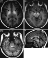
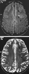
The purpose of this study was to describe the brain magnetic resonance imaging (MRI) findings in young patients with hepatosplenic schistosomiasis mansoni without overt neurologic manifestations. This study included 34 young persons (age range = 9-25 years) with hepatosplenic schistosomiasis mansoni who had been previously treated. Patients were scanned on a 1.5-T system that included multiplanar pre-contrast and post-contrast sequences, and reports were completed by two radiologists after a consensus review. Twenty (58.8%) patients had MRI signal changes that were believed to be related to schistosomiasis mansoni. Twelve of the 20 patients had small focal hyperintensities on T2WI in the cerebral white matter, and eight patients had symmetric hyperintense basal ganglia on T1WI. There was a high frequency of brain MRI signal abnormalities in this series. Although not specific, these findings may be related to schistosomiasis.
Figures


Similar articles
-
Chronic hepatosplenic schistosomiasis mansoni: magnetic resonance imaging and magnetic resonance angiography findings.Acta Radiol. 2007 Mar;48(2):125-34. doi: 10.1080/02841850601105833. Acta Radiol. 2007. PMID: 17354130
-
Evaluation of the cytokines IL-10 and IL-13 as mediators in the progression of Symmers fibrosis in patients with hepatosplenic schistosomiasis mansoni.Rev Col Bras Cir. 2010 Oct;37(5):333-7. doi: 10.1590/s0100-69912010000500005. Rev Col Bras Cir. 2010. PMID: 21180998 English, Portuguese.
-
Immunology of hepatosplenic schistosomiasis mansoni: a human perspective.Microbes Infect. 1999 Jun;1(7):553-60. doi: 10.1016/s1286-4579(99)80095-1. Microbes Infect. 1999. PMID: 10603572 Review. No abstract available.
-
Magnetic resonance imaging of the liver in hepatosplenic schistosomiasis mansoni.Rev Soc Bras Med Trop. 2002 Nov-Dec;35(6):679-80. doi: 10.1590/s0037-86822002000600022. Epub 2003 Feb 26. Rev Soc Bras Med Trop. 2002. PMID: 12612754 No abstract available.
-
Schistosoma mansoni: assessment of morbidity before and after control.Acta Trop. 2000 Oct 23;77(1):101-9. doi: 10.1016/s0001-706x(00)00124-8. Acta Trop. 2000. PMID: 10996126 Review.
Cited by
-
Diagnosis of coinfection by schistosomiasis and viral hepatitis B or C using 1H NMR-based metabonomics.PLoS One. 2017 Aug 1;12(8):e0182196. doi: 10.1371/journal.pone.0182196. eCollection 2017. PLoS One. 2017. PMID: 28763497 Free PMC article.
-
Magnetic Resonance Spectroscopy for Evaluating Portal-Systemic Encephalopathy in Patients with Chronic Hepatic Schistosomiasis Japonicum.PLoS Negl Trop Dis. 2016 Dec 15;10(12):e0005232. doi: 10.1371/journal.pntd.0005232. eCollection 2016 Dec. PLoS Negl Trop Dis. 2016. PMID: 27977668 Free PMC article.
-
Schistosomiasis: current epidemiology and management in travelers.Curr Infect Dis Rep. 2013 Jun;15(3):211-5. doi: 10.1007/s11908-013-0329-1. Curr Infect Dis Rep. 2013. PMID: 23568567
-
Case Report: Multiple Schistosomiasis Japonica Cerebral Granulomas without Gastrointestinal System Involvement: Report of Two Cases and Review of Literature.Am J Trop Med Hyg. 2020 Jun;102(6):1376-1381. doi: 10.4269/ajtmh.19-0797. Am J Trop Med Hyg. 2020. PMID: 32274982 Free PMC article. Review.
-
Increased IL-17, a Pathogenic Link between Hepatosplenic Schistosomiasis and Amyotrophic Lateral Sclerosis: A Hypothesis.Case Reports Immunol. 2014;2014:804761. doi: 10.1155/2014/804761. Epub 2014 Jul 23. Case Reports Immunol. 2014. PMID: 25379310 Free PMC article.
References
-
- Coura JR, Amaral RS. Epidemiological and control aspects of schistosomiasis in Brazilian endemic areas. Mem Inst Oswaldo Cruz. 2004;99:13–19. - PubMed
-
- Magalhães-Santos IF, Lemaire DC, Andrade-Filho AS, Queiroz AC, Carvalho OM, Carmo TM, Siqueira IC, Andrade DM, Rego MF, Guedes AP, Reis MG. Antibodies to Schistosoma mansoni in human cerebrospinal fluid. Am J Trop Med Hyg. 2003;68:294–298. - PubMed
-
- Betting LE, Pirani C, Queiroz LS, Damasceno BP, Cendes F. Seizures and cerebral schistosomiasis. Arch Neurol. 2005;62:1008–1010. - PubMed
-
- Carvalho OA, Carvalho CM, Bacelar AL, Andrade-Filho AS, Costa G, Fontes JB, Assis T. Clinical and cerebrospinal fluid (CSF) profile and CSF criteria for the diagnosis of spinal cord schistosomiasis. Arq Neuropsiquiatr. 2003;61:353–358. - PubMed
MeSH terms
LinkOut - more resources
Full Text Sources
Medical

