. 2012 Jun 19;109(25):9828-32.
doi: 10.1073/pnas.1203212109. Epub 2012 Jun 4.

Photochemically driven redox chemistry induces protocell membrane pearling and division

Affiliations

Photochemically driven redox chemistry induces protocell membrane pearling and division

Ting F Zhu et al. Proc Natl Acad Sci U S A. .

Abstract

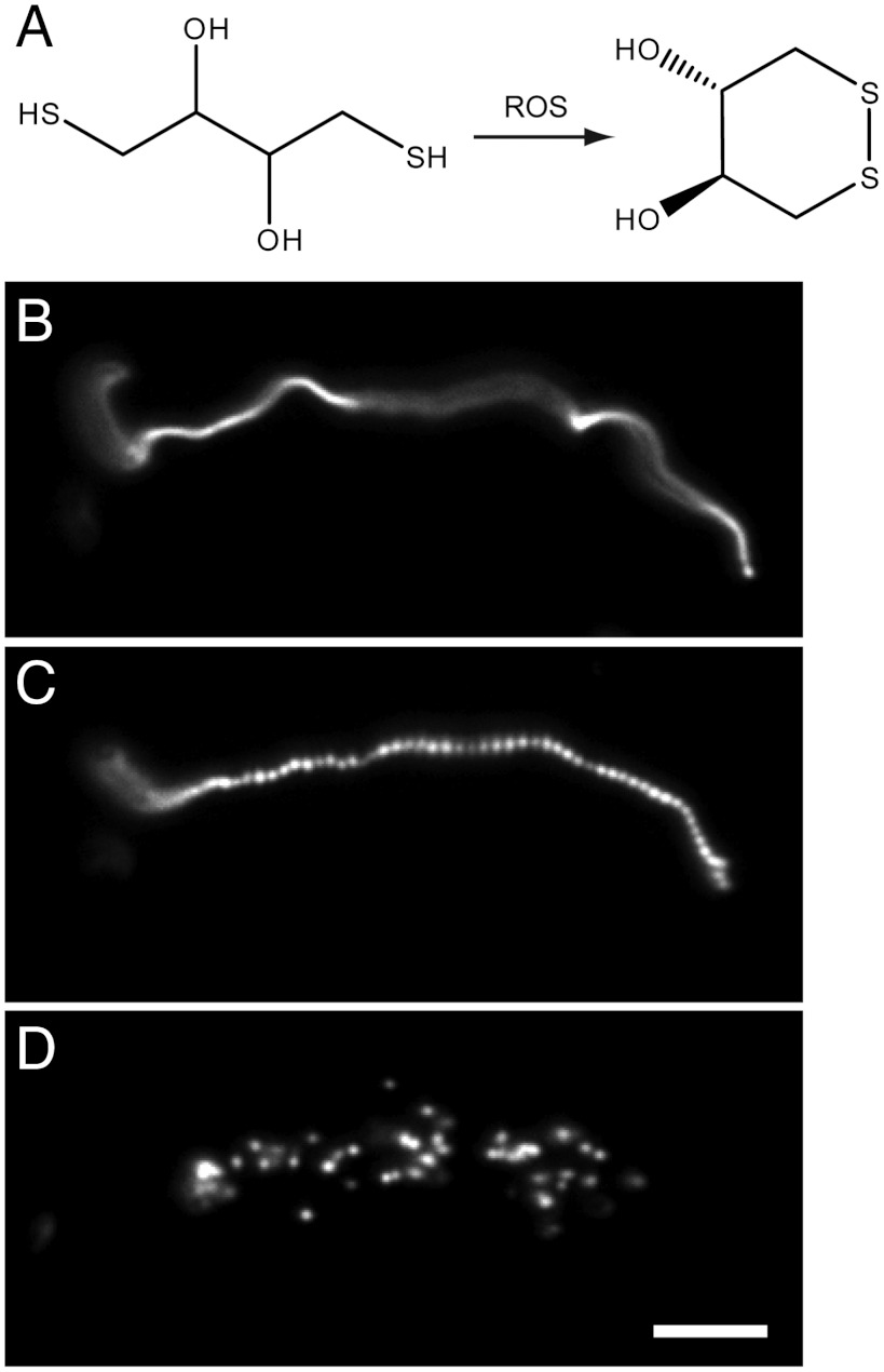
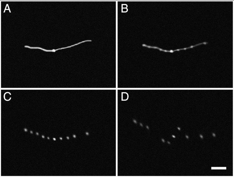
Prior to the evolution of complex biochemical machinery, the growth and division of simple primitive cells (protocells) must have been driven by environmental factors. We have previously demonstrated two pathways for fatty acid vesicle growth in which initially spherical vesicles grow into long filamentous vesicles; division is then mediated by fluid shear forces. Here we describe a different pathway for division that is independent of external mechanical forces. We show that the illumination of filamentous fatty acid vesicles containing either a fluorescent dye in the encapsulated aqueous phase, or hydroxypyrene in the membrane, rapidly induces pearling and subsequent division in the presence of thiols. The mechanism of this photochemically driven pathway most likely involves the generation of reactive oxygen species, which oxidize thiols to disulfide-containing compounds that associate with fatty acid membranes, inducing a change in surface tension and causing pearling and subsequent division. This vesicle division pathway provides an alternative route for the emergence of early self-replicating cell-like structures, particularly in thiol-rich surface environments where UV-absorbing polycyclic aromatic hydrocarbons (PAHs) could have facilitated protocell division. The subsequent evolution of cellular metabolic processes controlling the thiol:disulfide redox state would have enabled autonomous cellular control of the timing of cell division, a major step in the origin of cellular life.


Conflict of interest statement

The authors declare no conflict of interest.

Figures

Fig. 1.
Fig. 1.
Oleate vesicle pearling and division. (A) Radical-mediated oxidation of DTT. (B) An oleate vesicle (containing 2 mM HPTS, in 0.2 M Na-glycinamide, pH 8.5, 10 mM DTT) 30 min after the addition of five equivalents of oleate micelles. (C and D) Under intense illumination (for 2 s and 12 s, respectively), the long thread-like vesicle went through pearling and division (Movie S2). Scale bar, 10 μm.
Fig. 2.
Fig. 2.
NMR experiments to characterize the association of oxidized but not reduced DTT with oleate bilayer membranes. (A) Reference spectrum: conventional 1D 1H spectrum of oleic acid vesicles (15 mM oleate, 7.5 mM NaOD in 100% D2O, pH approximately 8.5), with 15 mM DTT and 15 mM trans-4,5-dihydroxy-1,2-dithiane added. Peaks corresponding to DTT (solid triangles) and its oxidized form trans-4,5-dihydroxy-1,2-dithiane (ox-DTT; stars) were assigned by spiking with the two compounds respectively. (B) STD (saturation transfer difference) spectrum of the above mixture in 100% D2O. The vinyl protons of oleic acid were selectively irradiated. Positive peaks reveal the interaction of trans-4,5-dihydroxy-1,2-dithiane molecules with oleate molecules of the bilayer membrane (stars). The absence of detectable peaks for DTT molecules suggests that DTT does not interact significantly with the oleate membranes. (C) WaterLOGSY (water ligand observed via gradient spectroscopy) spectrum of the above mixture in 10% D2O. Positive peaks show the interaction of oxidized DTT molecules with the oleate bilayer membranes (stars), while negative peaks suggest the absence of DTT association with bilayer membranes (solid triangles).
Fig. 3.
Fig. 3.
Oleate vesicle pearling and division with various thiols in the solution. (A) 3-mercaptopropionic acid. (B and C) An oleate vesicle (containing 2 mM HPTS, in 0.2 M Na-bicine, pH 8.5, 10 mM 3-mercaptopropionic acid, 30 min after the addition of five equivalents of oleate micelles) went through pearling and division under intense illumination (for 3 s and 15 s, respectively). (D) 3-mercapto-1-propanol. (E and F) An oleate vesicle as above but in 50 mM 3-mercapto-1-propanol, went through pearling and division under intense illumination (for 2 s and 10 s, respectively). (G) 1-mercapto-2-propanol. (H and I) An oleate vesicle as above but in 50 mM 1-mercapto-2-propanol went through pearling and division under intense illumination (for 2 s and 9 s, respectively). (J) 3-mercapto-1,2,4-triazole. (K and L) An oleate vesicle as above but in 50 mM 3-mercapto-1,2,4-triazole went through pearling and division under intense illumination (for 3 s and 13 s, respectively). Scale bar, 20 μm.
Fig. 4.
Fig. 4.
Oleate vesicle pearling and division mediated by the dipeptide di-L-cysteine. (A) An oleate vesicle (containing 2 mM HPTS, in 0.2 M Na-bicine, pH 8.5, 20 mM di-L-cysteine) 30 min after the addition of five equivalents of oleate micelles. (BD) Under intense illumination (for 3 s, 8 s, and 12 s, respectively), the long thread-like vesicle went through pearling and division. Scale bar, 10 μm.
Fig. 5.
Fig. 5.
Pearling and division of an oleate vesicle containing 1-hydroxypyrene in the membrane. (A) An oleate vesicle (with 20 mol%1-hydroxypyrene and 1 mol% Rh-DHPE in the membrane, in 0.2 M Na-bicine, pH 8.5, 15 mM DTT) 30 min after the addition of five equivalents of oleate micelles. (BD) Under intense illumination (for 57 s, 153 s, and 213 s, respectively), the long thread-like vesicle went through pearling and division (Movie S5). Scale bar, 10 μm.

References

    1. Szostak JW, Bartel DP, Luisi PL. Synthesizing life. Nature. 2001;409:387–390. - PubMed
    1. Budin I, Szostak JW. Physical effects underlying the transition from primitive to modern cell membranes. Proc Natl Acad Sci USA. 2011;108:5249–5254. - PMC - PubMed
    1. Chen IA, Roberts RW, Szostak JW. The emergence of competition between model protocells. Science. 2004;305:1474–1476. - PMC - PubMed
    1. Mansy SS, et al. Template-directed synthesis of a genetic polymer in a model protocell. Nature. 2008;454:122–125. - PMC - PubMed
    1. Hanczyc MM, Fujikawa SM, Szostak JW. Experimental models of primitive cellular compartments: Encapsulation, growth, and division. Science. 2003;302:618–622. - PMC - PubMed

Publication types