. 2012 Jun;85(1014):e195-205.
doi: 10.1259/bjr/78413721.

Primary and secondary breast lymphoma: prevalence, clinical signs and radiological features

Affiliations

Primary and secondary breast lymphoma: prevalence, clinical signs and radiological features

A Surov et al. Br J Radiol. 2012 Jun.

Abstract

Objectives: The purpose of this study was to determine the prevalence, clinical signs and radiological features of breast lymphoma.

Methods: This is a retrospective review of 36 patients with breast lymphoma (22 primary and 14 secondary). 35 patients were female and 1 was male; their median age was 65 years (range 24-88 years). In all patients, the diagnosis was confirmed histopathologically.

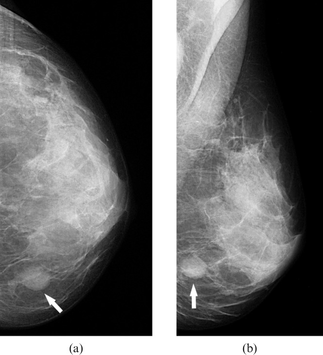
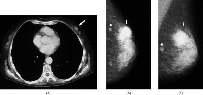
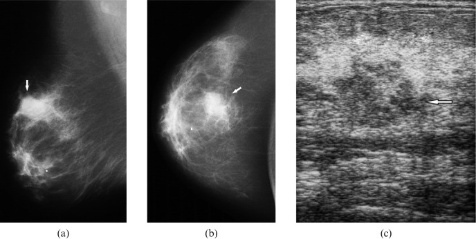
Results: The prevalence of breast lymphoma was 1.6% of all identified cases with non-Hodgkin lymphoma and 0.5% of cases with breast cancer. B-cell lymphoma was found in 94% and T-cell lymphoma in 6%. 96 lesions were identified (2.7 per patient). The mean size was 15.8 ± 8.3 mm. The number of intramammary lesions was higher in secondary than in primary lymphoma. The size of the identified intramammary lesions was larger in primary than in secondary lymphoma. Clinically, 86% of the patients presented with solitary or multiple breast lumps. In 14%, breast involvement was diagnosed incidentally during staging examinations.

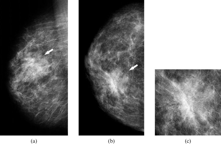
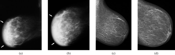
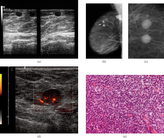
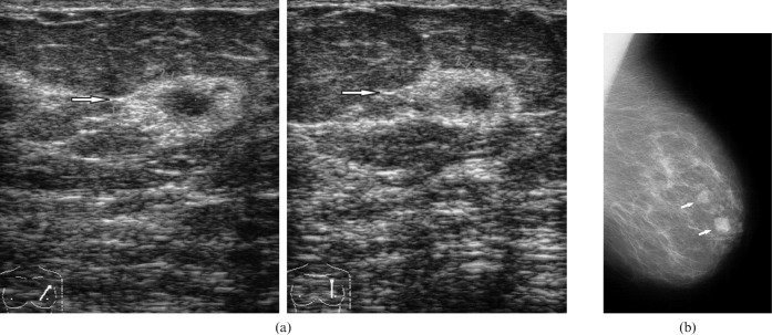
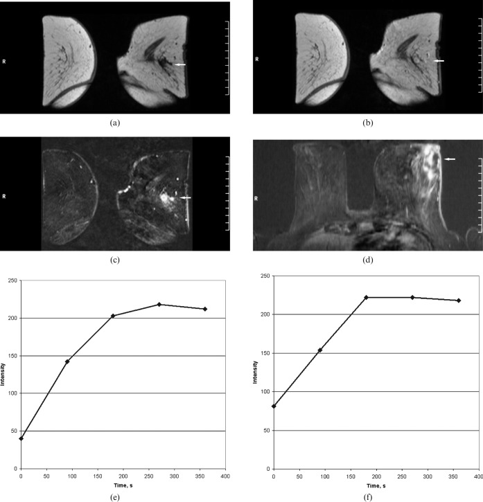
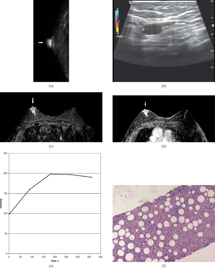
Conclusion: On mammography, intramammary masses were the most commonly seen (27 patients, 82%). Architectural distortion occurred in three patients (9%). In three patients (9%), no abnormalities were found on mammography. On ultrasound, the identified lesions were homogeneously hypoechoic or heterogeneously mixed hypo- to hyperechoic. On MRI, the morphology of the lesions was variable. After intravenous administration of contrast medium, a marked inhomogeneous contrast enhancement was seen in most cases. On CT, most lesions presented as circumscribed round or oval masses with moderate or high enhancement.


Figures

Figure 1
Figure 1
Craniocaudal (a) and mediolateral oblique mammograms (b) of the left breast in a patient with primary breast lymphoma showing a round, dense mass with circumscribed margins in the lower inner quadrant (arrows).
Figure 2
Figure 2
Radiological findings in a 62-year-old patient with secondary breast lymphoma. (a) CT of the chest documenting an irregular mass in the left breast (arrow). Craniocaudal (b) and mediolateral oblique mammograms (c) of the left breast showing a round, dense mass with irregular defined margins in the upper outer quadrant (small arrows). A small, well-defined mass in this quadrant is also seen (fat arrows).
Figure 3
Figure 3
Radiological images in a 59-year-old patient with a right breast lump and known history of primary lymphoma of the left breast, 22 years before this presentation. Mediolateral oblique (a) and craniocaudal mammograms (b) of the right breast showing a microlobulated dense mass in the upper outer quadrant (arrows). (c) On ultrasound, the mass was mixed hypo- to hyperechoic with indistinct margins (arrow).
Figure 4
Figure 4
Mediolateral oblique (a) and craniocaudal mammograms (b) of the right breast in a patient with primary breast lymphoma showing an asymmetric, poorly defined density containing architectural distortion (arrows) in the upper medial quadrant suspicious for a breast carcinoma. (c) Magnification view of the lesion.
Figure 5
Figure 5
Mammographic findings in a 68-year-old patient with secondary breast lymphoma. Mediolateral oblique (a) and craniocaudal mammograms (b) of the right breast showing a global asymmetry (diffuse opacity) of the parenchyma (arrows) with skin oedema compared with the left breast (c, d).
Figure 6
Figure 6
Breast images of a 64-year-old patient with secondary breast lymphoma. (a) Ultrasound showing circumscribed round anechoic lesions without posterior acoustic phenomena (arrows). (b) On mammography, the identified lesions were also well defined (arrow). Additionally, axillary lymph nodes were seen. (c) Magnification view of the lesion. (d) Power Doppler documenting the hypervascularity of one lesion. (e) Histological analysis after ultrasound-guided biopsy confirmed a B-cell lymphoma (Haematoxylin and eosin stain, ×100).
Figure 7
Figure 7
Radiological findings in a 77-year-old female with a left breast lump. (a) Ultrasound demonstrating a hypoechoic lesion (arrows) with a broad hyperechoic boundary and indistinct margins without posterior acoustic phenomenon. (b) Mammography showing dense lesions with circumscribed margins (arrows).
Figure 8
Figure 8
MRI of the breasts in a 61-year-old female with fever, weight loss and left breast swelling. (a) T1 weighted image before contrast administration confirmed masses in the left breast. These are isointense vs breast tissue (arrow). (b) First contrast-enhanced image showed a rapid contrast enhancement (arrow). (c) Subtracted images demonstrated multiple round lesions with marked enhancement. Additionally, a lateral cutaneous thickness of the left breast was seen (arrow). (d) Transverse reconstructions of a contrast-enhanced T1 weighted image showed additionally a moderate ductal enhancement between the lesions (arrow). (e, f) Time–intensity curves of the lesions. A strong rapid enhancement in the first minute associated with plateau (Type II curves). Time to peak 180 and 270 s; initial signal intensity, 174% and 407%.
Figure 9
Figure 9
Imaging findings in an 85-year-old male with a right breast lump. (a) On mammography, the identified lesion was oval in shape with circumscribed margins (arrow). (b) On ultrasound with power Doppler, the lesion was homogeneously anechoic with circumscribed margins and posterior acoustic enhancement. There was no vascularity. (c) T2 weighted image with fat saturation demonstrating a hyperintense lobulated lesion in the right breast (arrow). (d) Subtracted image after intravenous contrast administration showing a marked enhancement of the lesion (arrow). (e) Time–intensity curve of the main lesion. A strong rapid enhancement in the first minute associated with plateau (Type II curve). Time to peak, 180 s; initial signal intensity, 102%. (f) Histological examination after ultrasound-guided biopsy revealed a B-cell lymphoma (haematoxylin and eosin stain, ×200).

References

    1. Sabate JM, Gomez A, Torruba S, Camins A, Roson N, De LasHeras P, et al. Lymphoma of the breast: clinical and radiological features with pathologic correlation in 28 patients. Breast J 2002;8:294–304 - PubMed
    1. Giardini R, Piccolo C, Rilke F. Primary non-Hodgkin's lymphomas of the female breast. Cancer 1992;69:725–35 - PubMed
    1. Ribrag V, Bibeau F, El Weshi A, Frayfer J, Fadel C, Cebotaru C, et al. Primary breast lymphoma: a report of 20 cases. Br J Haematol 2001;115:253–6 - PubMed
    1. Hugh JC, Jackson FI, Hanson J, Poppema S. Primary breast lymphoma. An immunohistologic study of 20 new cases. Cancer 1990;66:2602–11 - PubMed
    1. Avilés A, Delgado S, Nambo MJ, Neri N, Murillo E, Cleto S. Primary breast lymphoma: results of a controlled clinical trial. Oncology 2005;69:256–60 - PubMed

MeSH terms