Review
. 2012 Aug 23;120(8):1552-61.
doi: 10.1182/blood-2012-04-408724. Epub 2012 Jun 4.

Does size matter in platelet production?

Affiliations
Review

Does size matter in platelet production?

Jonathan N Thon et al. Blood. .

Abstract

Platelet (PLT) production represents the final stage of megakaryocyte (MK) development. During differentiation, bone marrow MKs extend and release long, branched proPLTs into sinusoidal blood vessels, which undergo repeated abscissions to yield circulating PLTs. Circular-prePLTs are dynamic intermediate structures in this sequence that have the capacity to reversibly convert into barbell-proPLTs and may be related to "young PLTs" and "large PLTs" of both inherited and acquired macrothrombocytopenias. Conversion is regulated by the diameter and thickness of the peripheral microtubule coil, and PLTs are capable of enlarging in culture to generate barbell-proPLTs that divide to yield 2 smaller PLT products. Because PLT number and size are inversely proportional, this raises the question: do macrothrombocytopenias represent a failure in the intermediate stages of PLT production? This review aims to bring together and contextualize our current understanding of terminal PLT production against the backdrop of human macrothrombocytopenias to establish how "large PLTs" observed in both conditions are similar, how they are different, and what they can teach us about PLT formation. A better understanding of the cytoskeletal mechanisms that regulate PLT formation and determine PLT size offers the promise of improved therapies for clinical disorders of PLT production and an important source of PLTs for infusion.


Figures
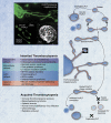
Figure 1
Figure 1
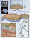
Cytoskeletal model of PLT production. Top left: immunofluorescence microscopy images of a proPLT producing MK, released barbell-proPLTs, and circular-prePLTs from a mouse fetal liver cell culture probed for β1-tubulin; rapid-freeze electron microscopy image of the PLT cytoskeleton. Bottom left: list of inherited thrombocytopenias affecting platelet size, grouped by underlying defect; list of common causes of acquired thrombocytopenia. Top right: model of the terminal stages of PLT production. Released proPLTs undergo successive rounds of fission along their midbody and at their ends to generate circular prePLTs and barbell-proPLTs. Circular prePLTs reversibly convert into barbell-proPLTs, from which PLTs are released after a final fission event at their midsection. PLTs may enlarge during culture and contribute to further PLT production. Bottom right: model of suspected errors in terminal PLT production that can account for phenotypes expressed in common inherited and acquired thrombocytopenias. For immunofluorescence microscopy, samples were fixed with 4% formaldehyde for 15 minutes and then permeabilized with 0.5% Triton X-100 and blocked in immunofluorescene blocking buffer (1% BSA, 0.05% sodium azide, and 10% FCS in PBS) overnight before antibody labeling. To demarcate permeabilized cells, samples were incubated with a rabbit polyclonal primary antibody for mouse tubulin generated against the C-terminal peptide sequence LEDSEEDAEEAEVEAEDKDH (Genemed Synthesis). Samples were treated with a secondary goat anti–rabbit antibody conjugated to an Alexa Fluor 488 (Invitrogen). Samples were examined with an Axiovert 200 microscope (Carl Zeiss) equipped with a 63× (numeric aperture, 1.4) PlanApoChromat oil immersion objective, and images were obtained using a charged coupled device camera (Hamamatsu). Images were analyzed using Metamorph image analysis Version 7.7.2.0 software (Molecular Devices) and ImageJ Version 1.45r software. Professional illustration by Alice Y. Chen.
Figure 2
Figure 2
Regulatory factors governing PLT formation and size. Top left: rapid-freeze electron microscopy image and illustration of major cytoskeletal components in the barbell-proPLT end. Boxed regions highlight specific regulatory factors governing PLT formation and size. For rapid-freeze electron microscopy, cells were placed in a solution of 0.75% Triton X-100 in piperazine-N,N-bis-2-ethanesulfonic acid, N-2-hydroxyethylpiperazine-N′-2-ethanesulfonic acid, ethyleneglycoltetraacetic acid, and MgCl2 (PHEM) containing 0.1% glutaraldehyde, 5μM phalloidin, and 30μM taxol, and attached to the surface of poly-L-lysine–coated coverslips by centrifugation at 280g for 5 minutes. The cytoskeleton was rinsed in PHEM solution and fixed for 15 minutes in 1% glutaraldehyde in PHEM. Coverslips were washed in distilled water, rapidly frozen in a liquid helium–cooled copper block, transferred to a liquid nitrogen–cooled stage, freeze-dried at −90°C, and metal cast with 1.2 nm of tantalum-tungsten with rotation at 45° and 3 nm of carbon at 90° without rotation. Replicas were floated, picked up on formvar-carbon–coated grid, and examined in a JEOL 1200-EX transmission electron microscope at 80 kV. Professional illustration by Alice Y. Chen.

References

    1. Italiano JE, Jr, Lecine P, Shivdasani RA, Hartwig JH. Blood platelets are assembled principally at the ends of proplatelet processes produced by differentiated megakaryocytes. J Cell Biol. 1999;147(6):1299–1312. - PMC - PubMed
    1. Kessel RG, Kardon RH. Circulating blood, blood vessels, and bone marrow. In: Johnson D, editor. Tissues and Organs: A Text-Atlas of Scanning Electron Microscopy. San Francisco, CA: W.H. Freeman; 1979. pp. 35–50.
    1. Behnke O. An electron microscope study of the rat megacaryocyte: II. Some aspects of platelet release and microtubules. J Ultrastruct Res. 1969;26(1):111–129. - PubMed
    1. Becker RP, De Bruyn PP. The transmural passage of blood cells into myeloid sinusoids and the entry of platelets into the sinusoidal circulation: a scanning electron microscopic investigation. Am J Anat. 1976;145(2):183–205. - PubMed
    1. Larson MK, Watson SP. A product of their environment: do megakaryocytes rely on extracellular cues for proplatelet formation? Platelets. 2006;17(7):435–440. - PubMed

Publication types